Copyright
©The Author(s) 2015.
World J Clin Cases. Mar 16, 2015; 3(3): 318-321
Published online Mar 16, 2015. doi: 10.12998/wjcc.v3.i3.318
Published online Mar 16, 2015. doi: 10.12998/wjcc.v3.i3.318
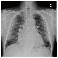
Figure 1 Chest X-ray showing mild cardiomegaly with prominence of central pulmonary arteries.
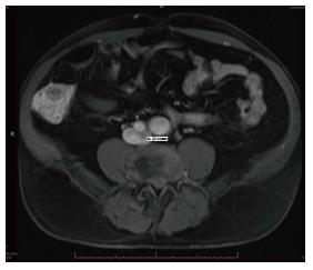
Figure 2 Magnetic resonance imaging of pelvis mark “A“ shows 21.
4 mm diameter of left common iliac artery.
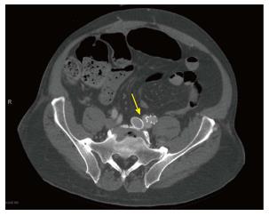
Figure 3 Computed tomography pelvis showing atriovenous fistula of left iliac vessels (yellow arrow) and stent in abdominal aorta.
- Citation: Singh S, Singh S, Jyothimallika J, Lynch TJ. May-Thurner syndrome: High output cardiac failure as a result of iatrogenic iliac fistula. World J Clin Cases 2015; 3(3): 318-321
- URL: https://www.wjgnet.com/2307-8960/full/v3/i3/318.htm
- DOI: https://dx.doi.org/10.12998/wjcc.v3.i3.318


