Copyright
©The Author(s) 2015.
World J Clin Cases. Mar 16, 2015; 3(3): 245-264
Published online Mar 16, 2015. doi: 10.12998/wjcc.v3.i3.245
Published online Mar 16, 2015. doi: 10.12998/wjcc.v3.i3.245
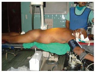
Figure 1 Position of patient.
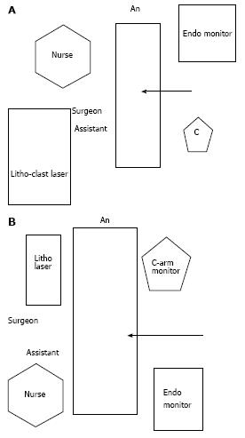
Figure 2 Arrangement of trolleys.
A: Arrangement for lower pole puncture; B: Arrangement for upper pole puncture.
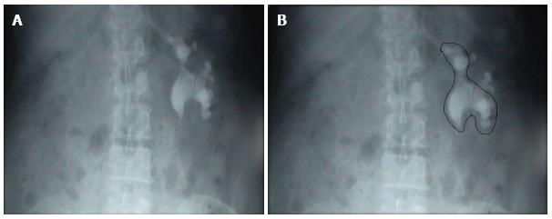
Figure 3 Outline-o-gram.
A: KUB showing a Left staghorn calculus; B: Outline-o-gram suggests that most of the calculus can be cleared by the lower puncture and a separate puncture may be needed for the residual fragment.
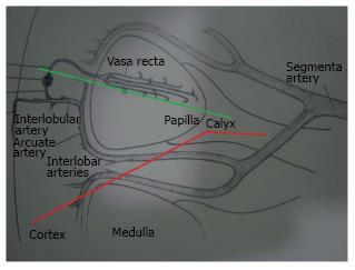
Figure 4 Arterial blood supply of kidney.
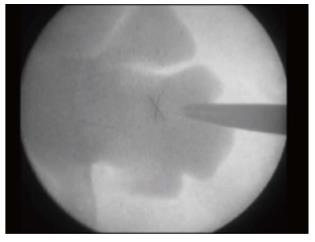
Figure 5 Trajectory of needle during puncture.
Figure 6 Bull’s eye appearance of the needle.
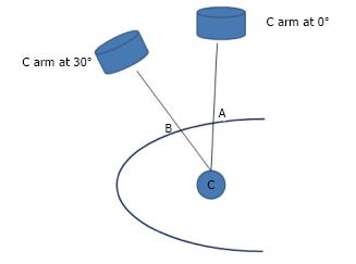
Figure 7 Hybrid technique.
Point C is the calyx to be punctured. Point A corresponds to the Point C with the C arm at 0°. Point B corresponds to the point C with the Carm rotated towards the surgeon by 30°. The needle held at Point A or B is seen as a Bull's eye effect on the Carm monitor. The distance between points A and B is measured.
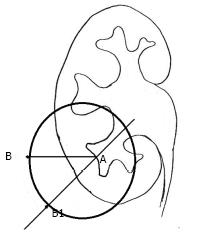
Figure 8 Hybrid technique.
“A” is the point on the skin which corresponds to the targeted calyx with the C arm at 0 degree and is the center of an imaginary circle. The distance between “A” to “B1” is equal to the distance between the “A” to “B”, i.e., the radius of the circle.
Figure 9 Hybrid technique.
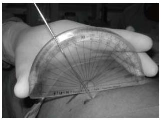
The angle which the needle makes with the skin surface is measured using a protractor.
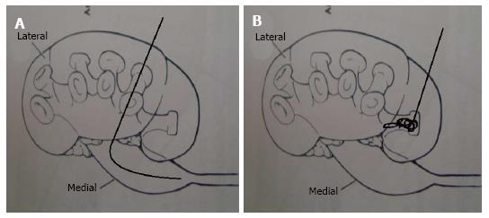
Figure 10 Calyceal puncture.
A: If the posterior calyx is punctured than the glide wire passes easily in the pelvis; B: if the anterior calyx is punctured than the glide wire gets coiled in the calyx before it makes its way to the pelvis.
- Citation: Sharma GR, Maheshwari PN, Sharma AG, Maheshwari RP, Heda RS, Maheshwari SP. Fluoroscopy guided percutaneous renal access in prone position. World J Clin Cases 2015; 3(3): 245-264
- URL: https://www.wjgnet.com/2307-8960/full/v3/i3/245.htm
- DOI: https://dx.doi.org/10.12998/wjcc.v3.i3.245


