Copyright
©The Author(s) 2015.
World J Clin Cases. Nov 16, 2015; 3(11): 951-955
Published online Nov 16, 2015. doi: 10.12998/wjcc.v3.i11.951
Published online Nov 16, 2015. doi: 10.12998/wjcc.v3.i11.951
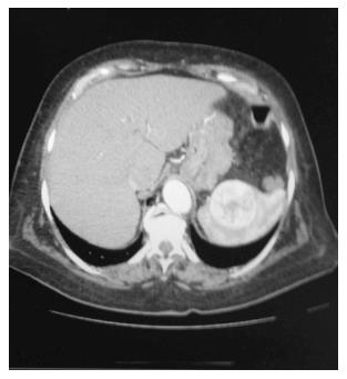
Figure 1 Computed tomography scan.
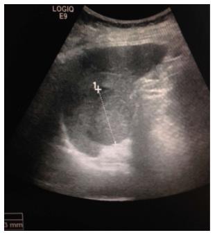
Figure 2 Ultrsound.
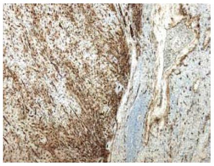
Figure 3 Positivity of vascular proliferation for immunohistochemical marker CD34; on right side there is normal splenic parenchyma (CD3450X).
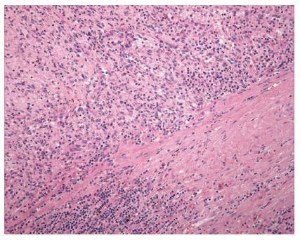
Figure 4 Vascular structures anastomosed (E-E 200 ×).
- Citation: Marzetti A, Messina F, Prando D, Verza LA, Vacca U, Azabdaftari A, Rubinato L, Reale D, Favat M, Barbujani M, Agresta F. Laparoscopic splenectomy for a littoral cell angioma of the spleen: Case report. World J Clin Cases 2015; 3(11): 951-955
- URL: https://www.wjgnet.com/2307-8960/full/v3/i11/951.htm
- DOI: https://dx.doi.org/10.12998/wjcc.v3.i11.951


