Copyright
©The Author(s) 2015.
World J Clin Cases. Nov 16, 2015; 3(11): 946-950
Published online Nov 16, 2015. doi: 10.12998/wjcc.v3.i11.946
Published online Nov 16, 2015. doi: 10.12998/wjcc.v3.i11.946
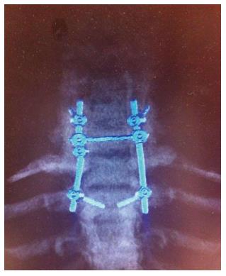
Figure 1 Maximum intensity projection reconstruction from computed tomography images shows C5-Th1 stabilization after the first intervention.
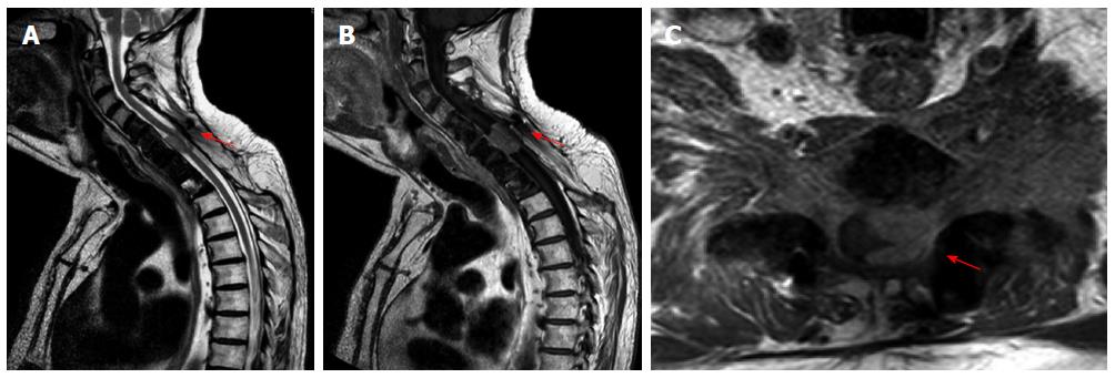
Figure 2 Cervical spine magnetic resonance imaging.
A: A C5-C7 lesion with homogeneous enhancement after gadolinium administration in the T1-weighted sequences; B: T2 weighted sequences showing the enclosed spinal cord; C: Especially on the left side (red arrow).
Figure 3 The immunohistochemical examination demonstrated positivity of neoplastic cells for (A) CK19, (B) p63 and (C) CD1a positive T-cells.
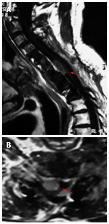
Figure 4 Magnetic resonance imaging of the cervico-thoracic spine at 3-mo follow-up showing (A) a small residual tumor, especially anteriorly to the cervical spinal cord and (B) axial view (red arrow).
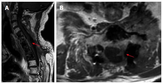
Figure 5 Cervical magnetic resonance imaging after radiation treatment showing (A) almost total disappearance of the mass and (B) axial view (red arrow).
- Citation: Marotta N, Mancarella C, Colistra D, Landi A, Dugoni DE, Delfini R. First description of cervical intradural thymoma metastasis. World J Clin Cases 2015; 3(11): 946-950
- URL: https://www.wjgnet.com/2307-8960/full/v3/i11/946.htm
- DOI: https://dx.doi.org/10.12998/wjcc.v3.i11.946


