Copyright
©The Author(s) 2015.
World J Clin Cases. Oct 16, 2015; 3(10): 920-925
Published online Oct 16, 2015. doi: 10.12998/wjcc.v3.i10.920
Published online Oct 16, 2015. doi: 10.12998/wjcc.v3.i10.920
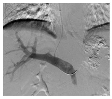
Figure 1 Direct portal vein radiography shows an enlarged portal vein; however, the splenic vein and gastric coronary vein could not be imaged.
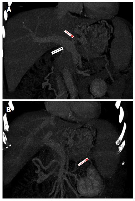
Figure 2 Enhanced computed tomography scan.
A: An enlarged portal vein from the origin of the gastric coronary vein and an enlarged, circuitous gastric coronary vein; B: Splenic vein flow signals in the portal venous phase are absent.
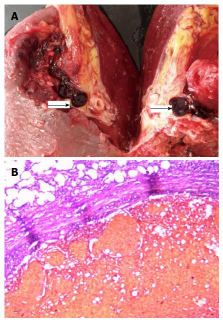
Figure 3 Anatomical resection of the spleen and hematoxylin-eosin staining staining.
A: The splenic vein is completely blocked by thrombosis; B: The splenic vein is completely filled by thrombosis.
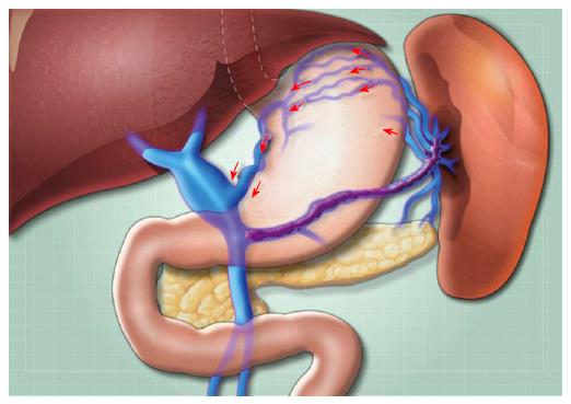
Figure 4 A schematic diagram of the pathophysiological and blood flow changes in this patient.
- Citation: Tang SH, Zeng WZ, He QW, Qin JP, Wu XL, Wang T, Wang Z, He X, Zhou XL, Fan QS, Jiang MD. Repeated pancreatitis-induced splenic vein thrombosis leads to intractable gastric variceal bleeding: A case report and review. World J Clin Cases 2015; 3(10): 920-925
- URL: https://www.wjgnet.com/2307-8960/full/v3/i10/920.htm
- DOI: https://dx.doi.org/10.12998/wjcc.v3.i10.920


