Copyright
©The Author(s) 2015.
World J Clin Cases. Oct 16, 2015; 3(10): 904-910
Published online Oct 16, 2015. doi: 10.12998/wjcc.v3.i10.904
Published online Oct 16, 2015. doi: 10.12998/wjcc.v3.i10.904
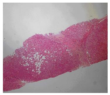
Figure 1 Liver biopsy showing an increased number of abnormal bile ducts, with nodularity of liver parenchyma accentuated by fibrous septa.
Figure 2 Eyes of the first patient.
Figure 3 Mouth of the first patient.
This appearance helps us to make a differential diagnosis of Cohen’s syndrome, in which a distinct cheerful facial expression is noted.
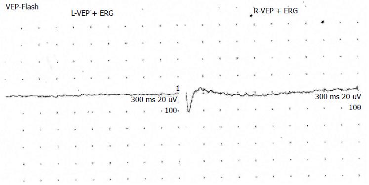
Figure 4 Electroretinography of case 3 showing no response in eyes bilaterally.
VEP: Visual evoked potantial; ERG: Electroretinography.
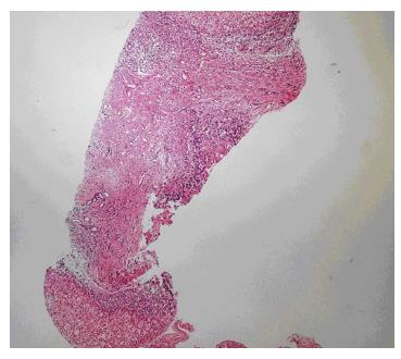
Figure 5 Liver showing nodular appearance due to fibrous bands in which elongated and angulated bile ducts are seen.
Figure 6 Liver showing nodular appearance due to fibrous bands in which bile ducts are elongated and periportal ductular proliferation is seen.
- Citation: Bayraktar Y, Yonem O, Varlı K, Taylan H, Shorbagi A, Sokmensuer C. Novel variant syndrome associated with congenital hepatic fibrosis. World J Clin Cases 2015; 3(10): 904-910
- URL: https://www.wjgnet.com/2307-8960/full/v3/i10/904.htm
- DOI: https://dx.doi.org/10.12998/wjcc.v3.i10.904


