Copyright
©The Author(s) 2015.
World J Clin Cases. Oct 16, 2015; 3(10): 894-899
Published online Oct 16, 2015. doi: 10.12998/wjcc.v3.i10.894
Published online Oct 16, 2015. doi: 10.12998/wjcc.v3.i10.894
Figure 1 Computed tomography abdomen and pelvis, axial view of hypodense splenic lesion.
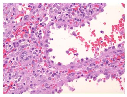
Figure 2 High power view of the tumor demonstrates tall columnar endothelial cells that line the cyst-like spaces.
These cells show no cytologic, nuclear atypia or mitotic figures (H and E stain, × 400).
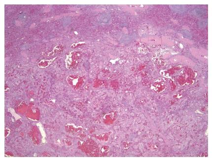
Figure 3 Low power view of the well-demarcated tumor with uninvolved spleen.
The tumor has anastomosing vascular channels and cyst-like hemorrhagic spaces.
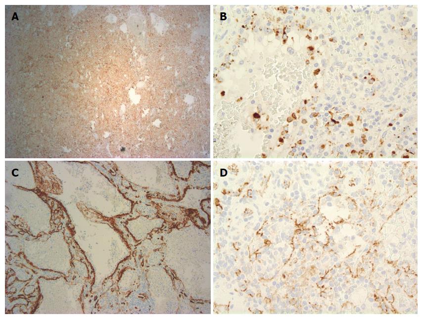
Figure 4 Endothelial cells lining the cyst-like spaces are immunoreactive.
A: CD68 (CD68 stain, × 100); B: Histiocytic marker lysozyme (lysozyme stain, × 400); C: Endothelial marker CD34 and the histocytoid cells are negative for CD34 (CD34 stain, × 400); D: Endothelial marker CD31 (CD31 stain, × 400).
- Citation: Bailey A, Vos J, Cardinal J. Littoral cell angioma: A case report. World J Clin Cases 2015; 3(10): 894-899
- URL: https://www.wjgnet.com/2307-8960/full/v3/i10/894.htm
- DOI: https://dx.doi.org/10.12998/wjcc.v3.i10.894


