Copyright
©The Author(s) 2015.
World J Clin Cases. Jan 16, 2015; 3(1): 34-41
Published online Jan 16, 2015. doi: 10.12998/wjcc.v3.i1.34
Published online Jan 16, 2015. doi: 10.12998/wjcc.v3.i1.34
Figure 1 Indications for enamel microabrasion.
Tooth staining from A: Fluorosis; B: Mineralized white spots.
Figure 2 Deep enamel staining due to hypoplasia.
A: Hypoplasia; B: Ineffective microabrasion treatment of the right central incisor.
Figure 3 Transillumination to determine staining depth.
A: Enamel hypoplasia in both central incisors; B, C: Transillumination to evaluate the staining.
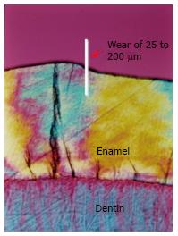
Figure 4 Depth of enamel removal.
Polarized light microscopy showing the ground tooth section after enamel microabrasion with Opalustre (reprinted with permission from Sundfeld et al[44]).
Figure 5 Enamel alterations after microabrasion.
Scanning electron microscopy showing the acid conditioning pattern on the enamel surface caused by microabrasion with A: Phosphoric acid and pumice; B: Opalustre; C: Confocal laser scanning microscopy showing the minimal alteration of the enamel surface, but intact subsurface, after microabrasion with Opalustre.
Figure 6 Resolution of fluorosis staining by microabrasion.
A: Clinical case of fluorosis before treatment; B: Results after enamel microabrasion (reprinted with permission Machado et al[58]).
- Citation: Pini NIP, Sundfeld-Neto D, Aguiar FHB, Sundfeld RH, Martins LRM, Lovadino JR, Lima DANL. Enamel microabrasion: An overview of clinical and scientific considerations. World J Clin Cases 2015; 3(1): 34-41
- URL: https://www.wjgnet.com/2307-8960/full/v3/i1/34.htm
- DOI: https://dx.doi.org/10.12998/wjcc.v3.i1.34


