Copyright
©2014 Baishideng Publishing Group Inc.
World J Clin Cases. Sep 16, 2014; 2(9): 482-487
Published online Sep 16, 2014. doi: 10.12998/wjcc.v2.i9.482
Published online Sep 16, 2014. doi: 10.12998/wjcc.v2.i9.482
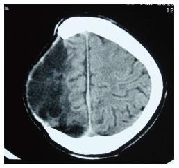
Figure 1 Computed tomography scan showing right cranial defect.
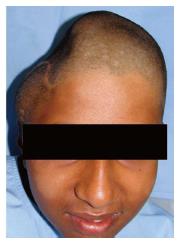
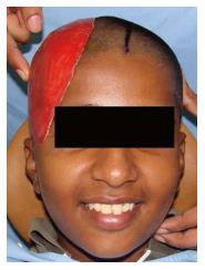
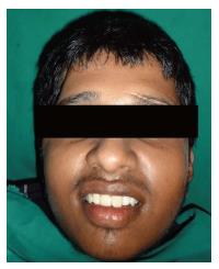
Figure 2 Frontal view of the cranial defect.
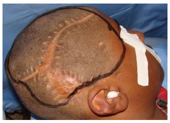
Figure 3 Patient preparation before impression making.
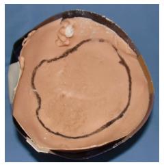
Figure 4 Tissue surface of the impression with defect marking.
Figure 5 Wax pattern try in.
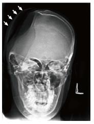
Figure 6 Radiographic verification of the wax pattern.
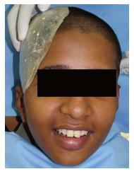
Figure 7 Try in of final heat cure alloplastic cranial implant.
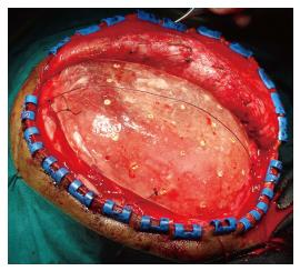
Figure 8 Placement and suturing of the final prosthesis.
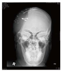
Figure 9 Post-operative postero-anterior view.
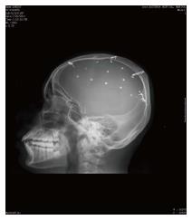
Figure 10 Post-operative lateral view.
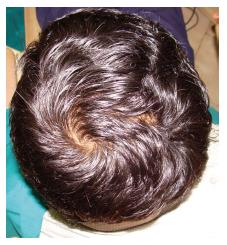
Figure 11 Restored cranial defect.
Figure 12 Frontal view of the restored cranial defect.
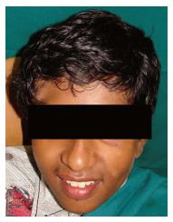
Figure 13 Follow up frontal profile.
- Citation: Gupta L, Aparna I, Balakrishnan D, Deenadayalan L, Hegde P, Agarwal P. Cranioplasty with custom made alloplastic prosthetic implant: A case report. World J Clin Cases 2014; 2(9): 482-487
- URL: https://www.wjgnet.com/2307-8960/full/v2/i9/482.htm
- DOI: https://dx.doi.org/10.12998/wjcc.v2.i9.482


