Copyright
©2014 Baishideng Publishing Group Inc.
World J Clin Cases. Sep 16, 2014; 2(9): 474-477
Published online Sep 16, 2014. doi: 10.12998/wjcc.v2.i9.474
Published online Sep 16, 2014. doi: 10.12998/wjcc.v2.i9.474
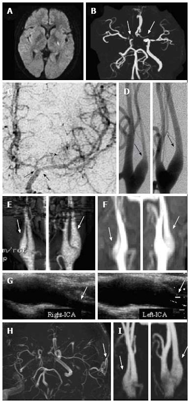
Figure 1 Imaging results.
A: Diffusion-weighted magnetic resonance imaging three hours after the onset of right hemiparesis revealed a hyperintense area in the posterior limb of the left internal capsule; B: Magnetic resonance angiography (MRA) showing bilateral severe stenosis of the distal portion of the internal carotid artery (ICA) and proximal portions of the middle cerebral artery (MCA) and anterior cerebral artery (ACA); C: Left carotid angiography (anteroposterior view), showing Moyamoya-like vascular changes around the circle of Willis (black dotted arrows) as well as stenosis of the distal left ICA and proximal left MCA and ACA (black arrows); Bilateral champagne bottle neck (CBN) signs at the proximal ICAs (arrows) were observed with D: Digital subtraction angiography; E: Three-dimensional computed tomography angiography; and F: Ultrasonography; G: MRA in the euthyroid state, showing no changes in the bilateral CBN signs (white arrows); H: MRA at four years after superficial temporal artery to MCA anastomosis, showing that the anastomosis remained patent (white arrow); I: No change was observed in the bilateral CBN signs after four years (white arrows)
- Citation: Shimogawa T, Morioka T, Sayama T, Hamamura T, Yasuda C, Arakawa S. Champagne bottle neck sign in a patient with Moyamoya syndrome. World J Clin Cases 2014; 2(9): 474-477
- URL: https://www.wjgnet.com/2307-8960/full/v2/i9/474.htm
- DOI: https://dx.doi.org/10.12998/wjcc.v2.i9.474


