Copyright
©2014 Baishideng Publishing Group Inc.
World J Clin Cases. Sep 16, 2014; 2(9): 455-458
Published online Sep 16, 2014. doi: 10.12998/wjcc.v2.i9.455
Published online Sep 16, 2014. doi: 10.12998/wjcc.v2.i9.455
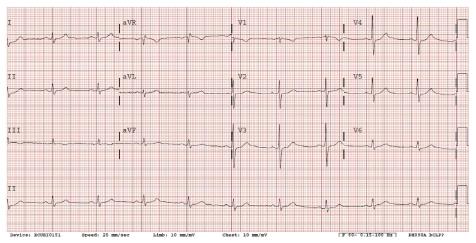
Figure 1 Electrocardiogram showed sinus bradycardia with a heart rate of 59 bpm and RSR’ in V1.
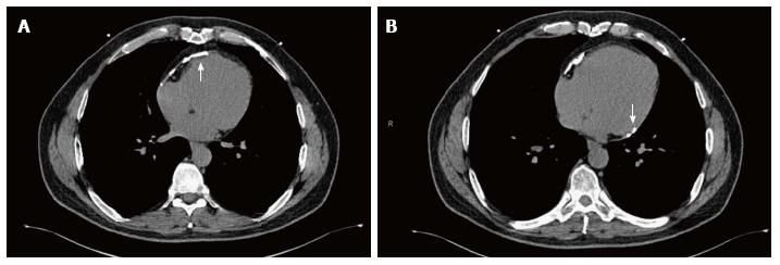
Figure 2 Non-contrast cardiac computed tomography for coronary artery calcium scoring showed moderate pericardial calcification mostly involving anterior (A) and inferobasal (B) portion of the pericardium.
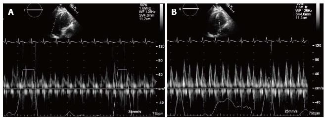
Figure 3 Apical four chamber view of transthoracic echocardiogram and pulse wave Doppler’s recording.
A: Tricuspid valve inflow in a patient with constrictive pericarditis; B: Mitral valve inflow in a patient with constrictive pericarditis. Note with inspiration, the E-wave velocity of mitral valve inflow decreases significantly. This reflects the hemodynamic changes in constrictive pericarditis, resulting from the lack of intrathoracic pressure transmission to the cardiac chambers and the exaggerated ventricular interdependence.
- Citation: Nguyen T, Phillips C, Movahed A. Incidental findings of pericardial calcification. World J Clin Cases 2014; 2(9): 455-458
- URL: https://www.wjgnet.com/2307-8960/full/v2/i9/455.htm
- DOI: https://dx.doi.org/10.12998/wjcc.v2.i9.455


