Copyright
©2014 Baishideng Publishing Group Inc.
World J Clin Cases. Jul 16, 2014; 2(7): 297-300
Published online Jul 16, 2014. doi: 10.12998/wjcc.v2.i7.297
Published online Jul 16, 2014. doi: 10.12998/wjcc.v2.i7.297
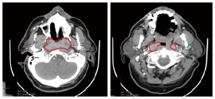
Figure 1 A large lesion arising from the posterior wall of the nasopharynx and bilateral lymphnode metastasis were detected in the parapharyngeal space (October 2009).
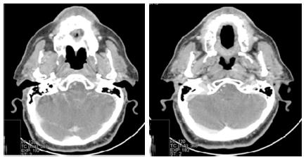
Figure 2 After neoadjuvant TPF followed by cetuximab-radiotherapy, a complete response was obtained (March 2010).
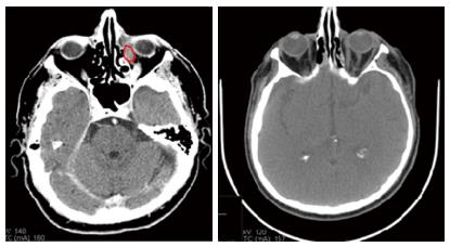
Figure 3 Computed tomography scan showed an intraorbital lesion (May 2012).
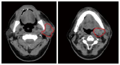
Figure 4 Computed tomography scan showed that the only site of persistent disease was the lymph node mass in the neck (December 2012).
- Citation: Perri F, Dell’Oca I, Muto P, Schiavone C, Aversa C, Fulciniti F, Solla R, Scarpati GDV, Buonerba C, Lorenzo GD, Caponigro F. Optimal management of a patient with recurrent nasopharyngeal carcinoma. World J Clin Cases 2014; 2(7): 297-300
- URL: https://www.wjgnet.com/2307-8960/full/v2/i7/297.htm
- DOI: https://dx.doi.org/10.12998/wjcc.v2.i7.297


