Copyright
©2014 Baishideng Publishing Group Inc.
World J Clin Cases. Jun 16, 2014; 2(6): 235-239
Published online Jun 16, 2014. doi: 10.12998/wjcc.v2.i6.235
Published online Jun 16, 2014. doi: 10.12998/wjcc.v2.i6.235
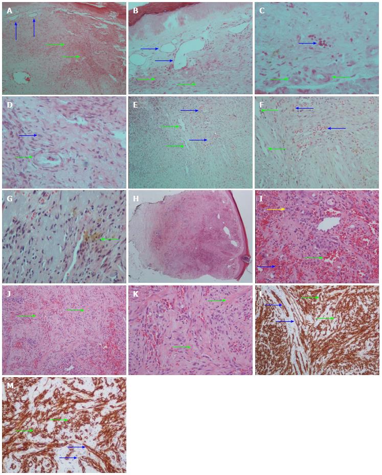
Figure 1 Microphotographs.
A, B: The first skin biopsy showing proliferation of fibroblasts (green arrows) and capillaries (blue arrows) in the dermis (H and E, A, × 100; B, × 200); C: The same biopsy showing narrowing of the lumen of medium size vessels (green arrow) and blood cell extravasation (blue arrow) (H and E, × 200); D: The same biopsy showing lumen narrowing of a medium size vessel (green arrows) and fibroblast proliferation (blue arrow) (H and E, × 400); E, F: The second skin biopsy showing dilated and irregular vessels (green arrows) and blood extravasation (blue arrows). (H and E, E, × 100; F, × 200); G: The same biopsy showing the presence of siderophages (green arrow) (H and E, × 400); H: The third skin biopsy showing a well-defined nodule composed of vascular spaces and spindle cells that has replaced the dermal collagen (H and E, × 20); I: At a higher magnification, there are blood-filled vessels (green arrow), spindle cells (yellow arrow) and blood cell extravasation (blue arrow) (H and E, × 200); J, K: The same biopsy showing compartmentalization of the nodule by bands of fibrocollagen tissue (green arrows); L, M: The same biopsy showing that spindle cells (green arrows) and endothelial vascular cells (blue arrows) expressed CD34. Streptavidin-biotin perixidase (L, × 100; M, ×200).
- Citation: Daoussis D, Chroni E, Tsamandas AC, Andonopoulos AP. Facial nerve palsy, headache, peripheral neuropathy and Kaposi’s sarcoma in an elderly man. World J Clin Cases 2014; 2(6): 235-239
- URL: https://www.wjgnet.com/2307-8960/full/v2/i6/235.htm
- DOI: https://dx.doi.org/10.12998/wjcc.v2.i6.235


