Copyright
©2014 Baishideng Publishing Group Inc.
World J Clin Cases. Jun 16, 2014; 2(6): 215-218
Published online Jun 16, 2014. doi: 10.12998/wjcc.v2.i6.215
Published online Jun 16, 2014. doi: 10.12998/wjcc.v2.i6.215
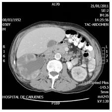
Figure 1 Contrast enhanced abdominal computed tomography shows a dilated and unstructured right kidney.
No mass effect images.
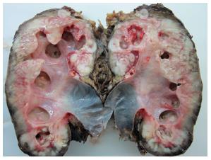
Figure 2 Macroscopic sagittal cut of the right kidney after nephrectomy, necrotic areas spread outside kidney and parenchyma is replaced by fibrotic tissue.
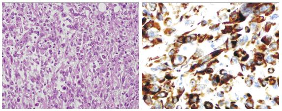
Figure 3 On the left side the sample (x 20) shows the invasive sarcomatoid pattern and on the right side the immunochemistry (x 40) positivity for CK7.
- Citation: Fernández-Pello S, Venta V, González I, Gil R, Menéndez CL. Pyonephrosis as a sign of sarcomatoid carcinoma of the renal pelvis. World J Clin Cases 2014; 2(6): 215-218
- URL: https://www.wjgnet.com/2307-8960/full/v2/i6/215.htm
- DOI: https://dx.doi.org/10.12998/wjcc.v2.i6.215


