Copyright
©2014 Baishideng Publishing Group Inc.
World J Clin Cases. Jun 16, 2014; 2(6): 206-208
Published online Jun 16, 2014. doi: 10.12998/wjcc.v2.i6.206
Published online Jun 16, 2014. doi: 10.12998/wjcc.v2.i6.206
Figure 1 Electrocardiogram.
Electrocardiogram on the top panel taken immediately after permanent pacemaker implantation demonstrates atrial sensing with ventricular pacing (A); The electrocardiogram on the bottom panel demonstrates failure to capture in the right ventricle with underlying 2:1 atrioventricular Block (B). The pacing configuration was bipolar with high outputs. Notice the prominent pacing spikes.
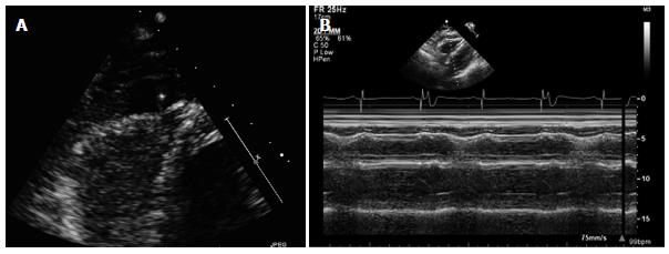
Figure 2 Transthoracic echocardiogram.
A: Image is an apical 4 chamber view of a transthoracic echocardiogram showing a moderate sized localized pericardial effusion with an echo bright structure within the effusion suspicious for lead perforation; B: Image is taken in M-mode and demonstrates right ventricular diastolic collapse suggestive of increased pericardial pressures.
Figure 3 This is an axial view of a non contrast chest computed tomography confirming diagnosis of right ventricular lead perforation.
The arrow points to the perforated lead. There is a pericardial effusion (PE). The right ventricle (RV) and left ventricle (LV) are also demonstrated.
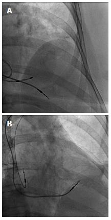
Figure 4 Image.
Top image shows the right ventricular permanent pacemaker lead protruding well past the heart border, A temporary transvenous pacemaker lead can also be seen within the right ventricle (A); The bottom image shows the repositioned right ventricular lead higher up on the interventricular septum and absence of the temporary transvenous pacemaker lead. The right atrial lead can also be seen in this image (B).
- Citation: Nash G, Williams JM, Nekkanti R, Movahed A. Case of early right ventricular pacing lead perforation and review of the literature. World J Clin Cases 2014; 2(6): 206-208
- URL: https://www.wjgnet.com/2307-8960/full/v2/i6/206.htm
- DOI: https://dx.doi.org/10.12998/wjcc.v2.i6.206


