Copyright
©2014 Baishideng Publishing Group Co.
World J Clin Cases. Mar 16, 2014; 2(3): 78-85
Published online Mar 16, 2014. doi: 10.12998/wjcc.v2.i3.78
Published online Mar 16, 2014. doi: 10.12998/wjcc.v2.i3.78
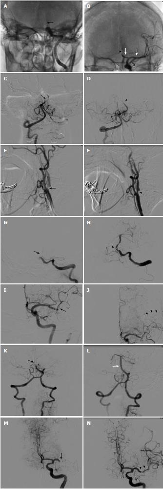
Figure 1 Case angiogram.
A: Pre-procedure imaging showed occlusion of distal left internal carotid artery (black arrow); B: Post-procedure imaging showed good flow of middle cerebral artery (MCA) and A1 (white arrow), occlusion of A2 (arrowhead) was noted; C: Pre-procedure imaging showed near total occlusion of mid basilar artery (BA) (black arrow); D: Post-procedure imaging showed improved in mid BA, distal BA (arrowhead) still occluded; E: Pre-procedure imaging showed critical stenosis of ostial left including internal carotid artery (ICA) (black arrow); F: Post-procedure imaging showed mild residual stenosis of proximal ICA after carotid stenting (arrowhead); G: Pre-procedure imaging showed proximal BA occlusion (black arrow); H: Post-procedure imaging showed patent BA with some residual thrombus in proximal part (arrowhead); I: Pre-procedure imaging showed thrombotic occlusion of superior M2 branch and slow flow of inferior M2 branch (black arrow); J: Post-procedure imaging showed good flow of inferior M2 branch, superior M2 branch still occluded and that area was supplied from pial collateral (arrowhead); K: Pre-procedure imaging showed occlusion of distal BA (black arrow); L: Post-procedure imaging showed patent BA (white arrow) and right posterior cerebral artery (PCA), with left PCA still occluded (arrowhead); M: Pre-procedure imaging showed occlusion of distal left M1 (black arrow); N: Post-procedure imaging showed good flow of left MCA (arrowhead) and all branches.
- Citation: Jongsathapongpan A, Raumthanthong A, Muengtaweepongsa S. Successful recanalization with multimodality endovascular interventional therapy in acute ischemic stroke. World J Clin Cases 2014; 2(3): 78-85
- URL: https://www.wjgnet.com/2307-8960/full/v2/i3/78.htm
- DOI: https://dx.doi.org/10.12998/wjcc.v2.i3.78


