Copyright
©2014 Baishideng Publishing Group Co.
World J Clin Cases. Feb 16, 2014; 2(2): 48-51
Published online Feb 16, 2014. doi: 10.12998/wjcc.v2.i2.48
Published online Feb 16, 2014. doi: 10.12998/wjcc.v2.i2.48
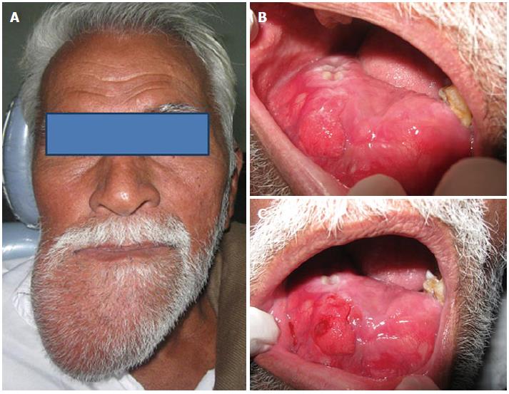
Figure 1 Extra (A) and intra oral photograph of swelling with labial, buccal and lingual cortical expansion (B, C).
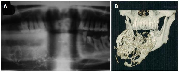
Figure 2 Orthopantomograph (A) and 3D computed tomography (B) showing honeycomb lesion.
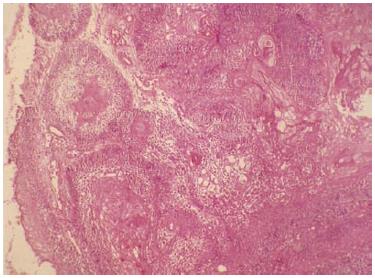
Figure 3 Histopathology specimen showing epithelial follicles with squamous metaplasia and numerous keratin pearls.
Figure 4 Post-op photograph (A) and orthopantomograph (B) of patient after surgical resection of the tumor by microvascular reconstructive surgery and reconstruction with a fibula graft.
- Citation: Srikanth MD, Radhika B, Metta K, Renuka NV. Ameloblastic carcinoma: Report of a rare case. World J Clin Cases 2014; 2(2): 48-51
- URL: https://www.wjgnet.com/2307-8960/full/v2/i2/48.htm
- DOI: https://dx.doi.org/10.12998/wjcc.v2.i2.48


