Copyright
©2014 Baishideng Publishing Group Co.
World J Clin Cases. Feb 16, 2014; 2(2): 42-44
Published online Feb 16, 2014. doi: 10.12998/wjcc.v2.i2.42
Published online Feb 16, 2014. doi: 10.12998/wjcc.v2.i2.42
Figure 1 Axial T1 weighted image (A), T2 weighted image (B) and contrast enhanced fat saturated T1 image (C) showing contrast enhancing inguinal conglomerated lymph node enlargement (arrows).
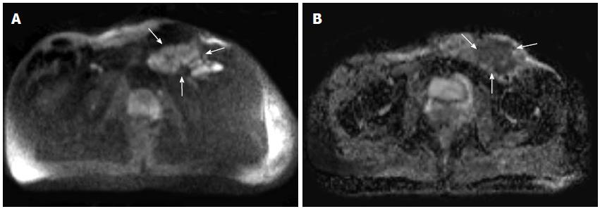
Figure 2 Diffusion weighted images b800 (A) and apparent diffusion coefficient map (B) showing diffusion restriction in inguinal conglomerated lymph nodes (arrows).
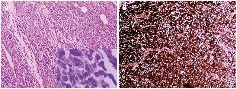
Figure 3 Pathological examination.
A: Photomicrograph of metastatic malignant melanoma in the inguinal lymph node (HE, × 50). The bottom right corner HE, ×100; B: Immunohistochemical study shows that the spindle cells are positive for HMB-45.
- Citation: Bayraktutan U, Kantarci M, Pirimoglu B, Ogul H, Okur A, Gursan N. Utility of diffusion-weighted imaging in the diagnosis of inguinal lymph node metastasis with malignant melanoma. World J Clin Cases 2014; 2(2): 42-44
- URL: https://www.wjgnet.com/2307-8960/full/v2/i2/42.htm
- DOI: https://dx.doi.org/10.12998/wjcc.v2.i2.42


