Copyright
©2014 Baishideng Publishing Group Co.
World J Clin Cases. Feb 16, 2014; 2(2): 36-38
Published online Feb 16, 2014. doi: 10.12998/wjcc.v2.i2.36
Published online Feb 16, 2014. doi: 10.12998/wjcc.v2.i2.36
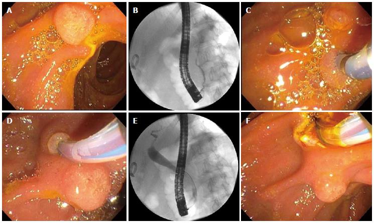
Figure 1 The patient underwent an endoscopic retrograde cholangiopancreatography for stone removal.
A: The major papilla appeared normal; B: Normal pancreatogram; C: The major papilla was re-examined; D: Cannulation of the bile duct through the second orifice; E: Biliary tract with biliary stone within the distal common bile duct; F: Biliary sphincterotomy.
- Citation: Chavalitdhamrong D, Draganov PV. Unexpected anomaly of the common bile duct and pancreatic duct. World J Clin Cases 2014; 2(2): 36-38
- URL: https://www.wjgnet.com/2307-8960/full/v2/i2/36.htm
- DOI: https://dx.doi.org/10.12998/wjcc.v2.i2.36


