Copyright
©2014 Baishideng Publishing Group Inc.
World J Clin Cases. Dec 16, 2014; 2(12): 927-929
Published online Dec 16, 2014. doi: 10.12998/wjcc.v2.i12.927
Published online Dec 16, 2014. doi: 10.12998/wjcc.v2.i12.927
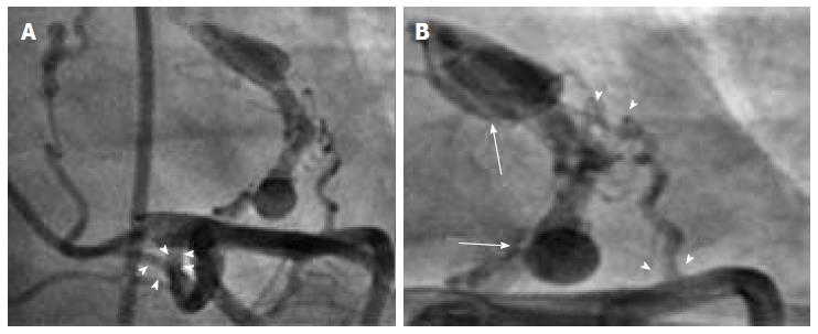
Figure 1 A: The fistula emerging from the left main coronary artery is divided into two branches after the fold (arrowhead); B: An angiographic view showing the combination of a small fistula emerging from left anterior descending artery, the fistula from left main coronary artery (arrowhead) and large saccular sacs on the fistula line (arrow).
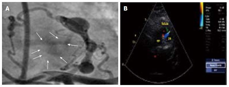
Figure 2 The passage of the contrast agent formed by the fistula (A), parasternal short axis echocardiography revealed the color flow associated with the fistula (B).
- Citation: Emre E, Aktas M, Sahin T, Ural E, Ural D. Rare multiple fistulas with large saccular aneurysms originating from left anterior descending artery and left main coronary artery. World J Clin Cases 2014; 2(12): 927-929
- URL: https://www.wjgnet.com/2307-8960/full/v2/i12/927.htm
- DOI: https://dx.doi.org/10.12998/wjcc.v2.i12.927


