Copyright
©2014 Baishideng Publishing Group Inc.
World J Clin Cases. Dec 16, 2014; 2(12): 918-923
Published online Dec 16, 2014. doi: 10.12998/wjcc.v2.i12.918
Published online Dec 16, 2014. doi: 10.12998/wjcc.v2.i12.918
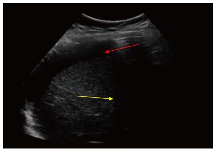
Figure 1 Abdominal ultrasound at epigastric level shows moderate amount of free fluid (yellow arrow) and reverberation artifact immediately under the abdominal wall compatible with intraperitoneal free air (red arrow).
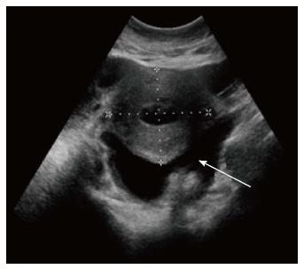
Figure 2 At pelvic level, enlarged uterus is observed associated with free fluid, which presents echogenic material within suggestive of pus (white arrow).
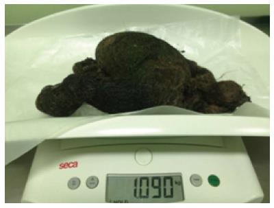
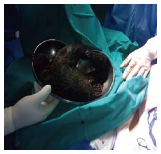
Figure 3 Trichobezoar extracted weighing 1.
09 kg.
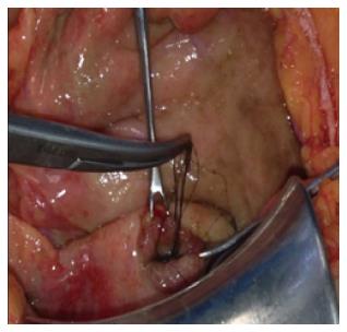
Figure 4 Finding of hair through perforated ulcer.
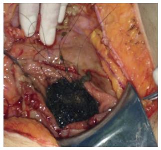
Figure 5 Gastric opening for extraction of trichobezoar.
Figure 6 Trichobezoar mass taking the shape of the stomach and part of the duodenum.
-
Citation: Morales HL, Catalán CH, Demetrio RA, Rivas ME, Parraguez NC, Alvarez MA. Gastric trichobezoar associated with perforated peptic ulcer and
Candida glabrata infection. World J Clin Cases 2014; 2(12): 918-923 - URL: https://www.wjgnet.com/2307-8960/full/v2/i12/918.htm
- DOI: https://dx.doi.org/10.12998/wjcc.v2.i12.918


