Copyright
©2014 Baishideng Publishing Group Inc.
World J Clin Cases. Dec 16, 2014; 2(12): 907-911
Published online Dec 16, 2014. doi: 10.12998/wjcc.v2.i12.907
Published online Dec 16, 2014. doi: 10.12998/wjcc.v2.i12.907
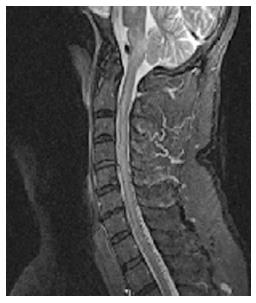
Figure 1 Magnetic resonance imaging of the cervical spine showing increased T2/STIR signal intensity in the pons, medulla, and upper cervical spine and multiple small flow voids in the dorsal cervicothoracic spine suggestive of dural arteriovenous malformation.
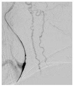
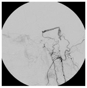
Figure 2 Angiogram showing a Cognard V tentorial dural arteriovenous fistula fed by the left middle meningeal, tentorial branch of the left ICA, and dural branches of the occipital and posterior auricular arteries, with drainage into cervical spinal veins.
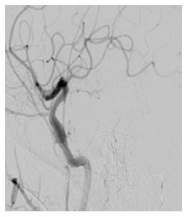
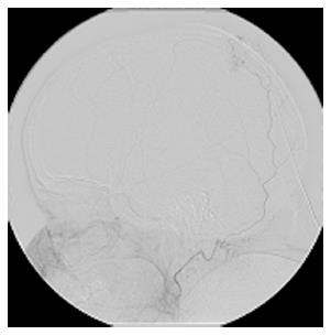
Figure 3 Arteriovenous fistula shown in Figure 2 now successfully embolized with Onyx without any residual filling seen following embolization.
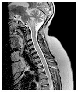
Figure 4 Magnetic resonance imaging brain and cervical spine showing asymmetric expansion and T2/FLAIR signal abnormality of the left side of the brainstem at the cervicomedullary junction, as well as diffuse expansion and mild diffuse T2 signal abnormality within the cervical and upper thoracic spinal cord.
Figure 5 Cerebral angiogram showed a left transverse sigmoid junction dural arteriovenous fistula fed by the left occipital artery.
Figure 6 Post onyx embolization angiogram shows no persistent opacification of the fistula with left occipital artery injection.
- Citation: Gross R, Ali R, Kole M, Dorbeistein C, Jayaraman MV, Khan M. Tentorial dural arteriovenous fistula presenting as myelopathy: Case series and review of literature. World J Clin Cases 2014; 2(12): 907-911
- URL: https://www.wjgnet.com/2307-8960/full/v2/i12/907.htm
- DOI: https://dx.doi.org/10.12998/wjcc.v2.i12.907


