Copyright
©2014 Baishideng Publishing Group Inc.
World J Clin Cases. Dec 16, 2014; 2(12): 903-906
Published online Dec 16, 2014. doi: 10.12998/wjcc.v2.i12.903
Published online Dec 16, 2014. doi: 10.12998/wjcc.v2.i12.903
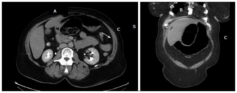
Figure 1 Computed tomography of abdomen and pelvis showing a loop of distended colon within lesser sac without bowel ischemia or perforation.
Radiological features include: (1) the cecum herniated (C) into the lesser sac behind the stomach (S) (A and B); (2) the presence of mesentery (white arrow) between the portal vein and inferior vena cava (A); and (3) the presence of gas or fluid in the lesser sac with its ‘beak’ directed toward the foramen of winslow (B).
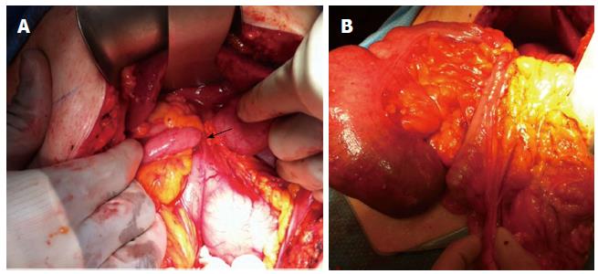
Figure 2 Intraoperatively, a cecal bascule was found herniating into lesser sac via foramen of winslow (A).
Upon reduction, the cecum appeared viable (B).
- Citation: Makarawo T, Macedo FI, Jacobs MJ. Cecal bascule herniation into the lesser sac. World J Clin Cases 2014; 2(12): 903-906
- URL: https://www.wjgnet.com/2307-8960/full/v2/i12/903.htm
- DOI: https://dx.doi.org/10.12998/wjcc.v2.i12.903


