Copyright
©2014 Baishideng Publishing Group Inc.
World J Clin Cases. Dec 16, 2014; 2(12): 899-902
Published online Dec 16, 2014. doi: 10.12998/wjcc.v2.i12.899
Published online Dec 16, 2014. doi: 10.12998/wjcc.v2.i12.899
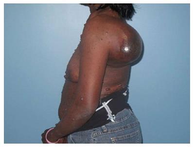
Figure 1 Left shoulder mass at initial presentation.
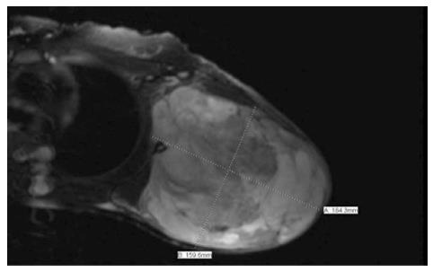
Figure 2 Magnetic resonance imaging of left shoulder, Mass: 18.
4 cm × 15.9 cm × 20 cm.
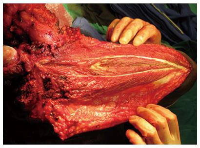
Figure 3 Fillet flap of the upper arm post subperiosteal dissection and removal of the humerus.
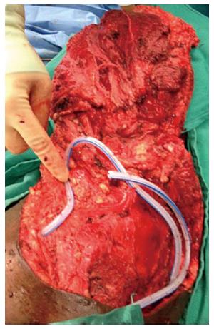
Figure 4 Fillet flap with drains, prior to closure.
- Citation: Singla P, Kachare SD, Fitzgerald TL, Zeri RS, Haque E. Reconstruction using a pedicled upper arm fillet flap after excision of a malignant peripheral nerve sheath tumor: A case report. World J Clin Cases 2014; 2(12): 899-902
- URL: https://www.wjgnet.com/2307-8960/full/v2/i12/899.htm
- DOI: https://dx.doi.org/10.12998/wjcc.v2.i12.899


