Copyright
©2014 Baishideng Publishing Group Inc.
World J Clin Cases. Dec 16, 2014; 2(12): 883-887
Published online Dec 16, 2014. doi: 10.12998/wjcc.v2.i12.883
Published online Dec 16, 2014. doi: 10.12998/wjcc.v2.i12.883
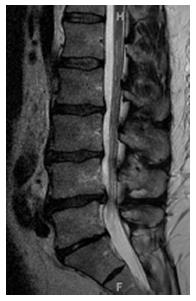
Figure 1 Mid-sagittal T2-weighted (3230, 120) image of the lumbar spine in a 50-year-old male with congenital lumbar spinal stenosis.
The lumbar spine shows loss of the lordotic curve, multilevel spondylolisthesis, and degenerative disc disease manifested as loss of disc height, circumferential disc bugles, anterior disc herniations, Schmorl’s modes and a central disc protrusion. In this subject, the mid-sagittal spinal canal diameter ranged from 1.45 cm at the L1 level to 1.03 cm at L4 level. The average mid-spinal canal diameter was 1.26 cm.
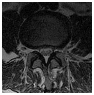
Figure 2 Axial T2-weighted (5000, 102) images of the lumbar spine in a 48-year-old female with congenital lumbar spinal stenosis exhibits a shallow annular disc bulge.
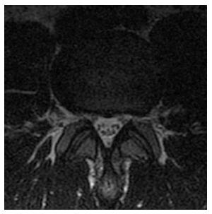
Figure 3 Axial T2-weighted (3516, 115) image of the lumbar spine in a 29-year-old male with congenital lumbar spinal stenosis demonstrates a circumferential disc bulge with a superimposed left foraminal disc protrusion.
- Citation: Soldatos T, Chalian M, Thawait S, Belzberg AJ, Eng J, Carrino JA, Chhabra A. Spectrum of magnetic resonance imaging findings in congenital lumbar spinal stenosis. World J Clin Cases 2014; 2(12): 883-887
- URL: https://www.wjgnet.com/2307-8960/full/v2/i12/883.htm
- DOI: https://dx.doi.org/10.12998/wjcc.v2.i12.883


