Copyright
©2014 Baishideng Publishing Group Inc.
World J Clin Cases. Nov 16, 2014; 2(11): 717-723
Published online Nov 16, 2014. doi: 10.12998/wjcc.v2.i11.717
Published online Nov 16, 2014. doi: 10.12998/wjcc.v2.i11.717
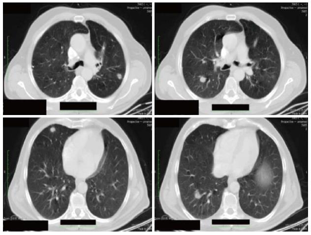
Figure 1 Thorax computed tomography-scan before starting chemotherapy (October 2008).
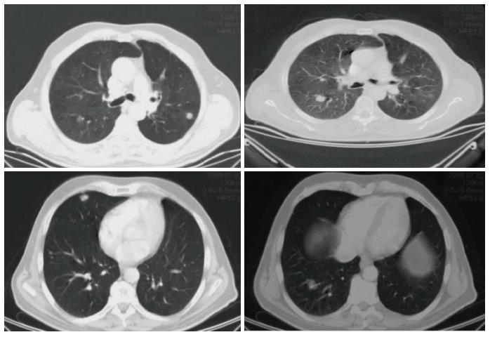
Figure 2 Thorax computed tomography-scan after 8 cycles of treatment.
Note stabilization of the disease according to RECIST criteria (June 2009).
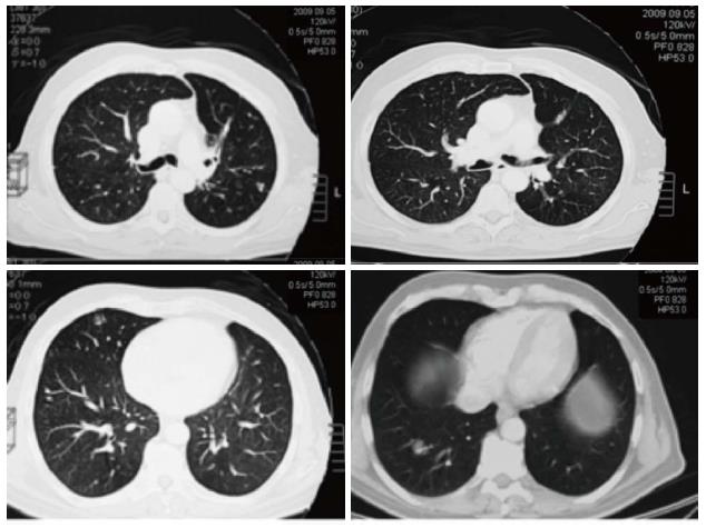
Figure 3 Thorax computed tomography-scan after two months of maintenance treatment.
Note a partial regression of disease according to RECIST criteria (September 2009).
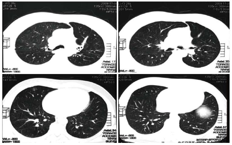
Figure 4 Thorax computed tomography-scan after 4 mo of maintenance treatment.
The disease has completely regressed according to RECIST criteria (November 2009).
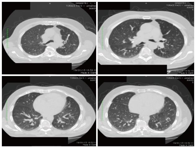
Figure 5 Thorax computed tomography-scan showing the complete response after discontinuing bevacizumab (January 2013).
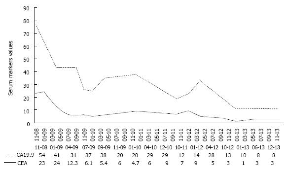
Figure 6 CEA (ng/mL) and CA19.
9 (U/mL) serum levels throughout the treatment and follow-up period.
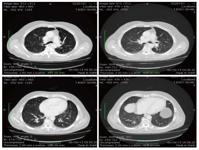
Figure 7 Thorax computed tomography-scan, showing the response at 60 mo since diagnosis (September 2013).
- Citation: Stefano AD, Moretto R, Cella CA, Romano FJ, Raimondo L, Fiore G, Pietro FD, Pepe S, Placido SD, Carlomagno C. Bevacizumab maintenance in metastatic colorectal cancer: How long? World J Clin Cases 2014; 2(11): 717-723
- URL: https://www.wjgnet.com/2307-8960/full/v2/i11/717.htm
- DOI: https://dx.doi.org/10.12998/wjcc.v2.i11.717


