Copyright
©2014 Baishideng Publishing Group Inc.
World J Clin Cases. Oct 16, 2014; 2(10): 591-595
Published online Oct 16, 2014. doi: 10.12998/wjcc.v2.i10.591
Published online Oct 16, 2014. doi: 10.12998/wjcc.v2.i10.591
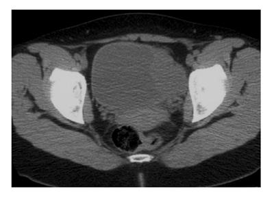
Figure 1 Computer tomography showing an avidly enhancing mass in the left urinary bladder wall.
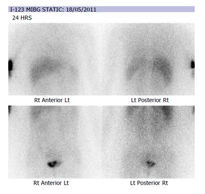
Figure 2 Iodine-123-meta-iodobenzylguanidine scintiscan showing intense tracer uptake in the left side of the bladder consistent with bladder paraganglioma as well as injection artefact in the right cubital fossa.
I-123 MIBG: Iodine-123-meta-iodobenzylguanidine.
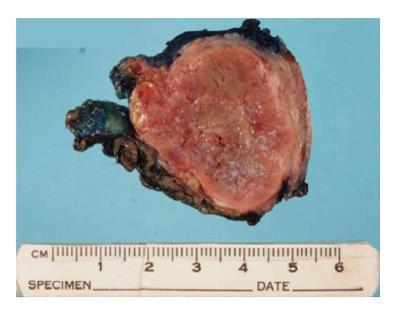
Figure 3 Macroscopic view of bladder paraganglioma.
The tumour is red-brown in colour and well-circumscribed.
Figure 4 Haematoxylin and eosin staining of bladder paraganglioma tumour (A) showing characteristic nests of cells with eosinophilic cytoplasm and round nuclei.
Chromogranin staining (B) was strongly positive, whilst S100 staining (C) was positive in patches.
Figure 5 Longitudinal and transverse views of a paraganglioma protruding into the bladder.
- Citation: Ranaweera M, Chung E. Bladder paraganglioma: A report of case series and critical review of current literature. World J Clin Cases 2014; 2(10): 591-595
- URL: https://www.wjgnet.com/2307-8960/full/v2/i10/591.htm
- DOI: https://dx.doi.org/10.12998/wjcc.v2.i10.591


