Copyright
©The Author(s) 2025.
World J Clin Cases. Mar 26, 2025; 13(9): 96897
Published online Mar 26, 2025. doi: 10.12998/wjcc.v13.i9.96897
Published online Mar 26, 2025. doi: 10.12998/wjcc.v13.i9.96897
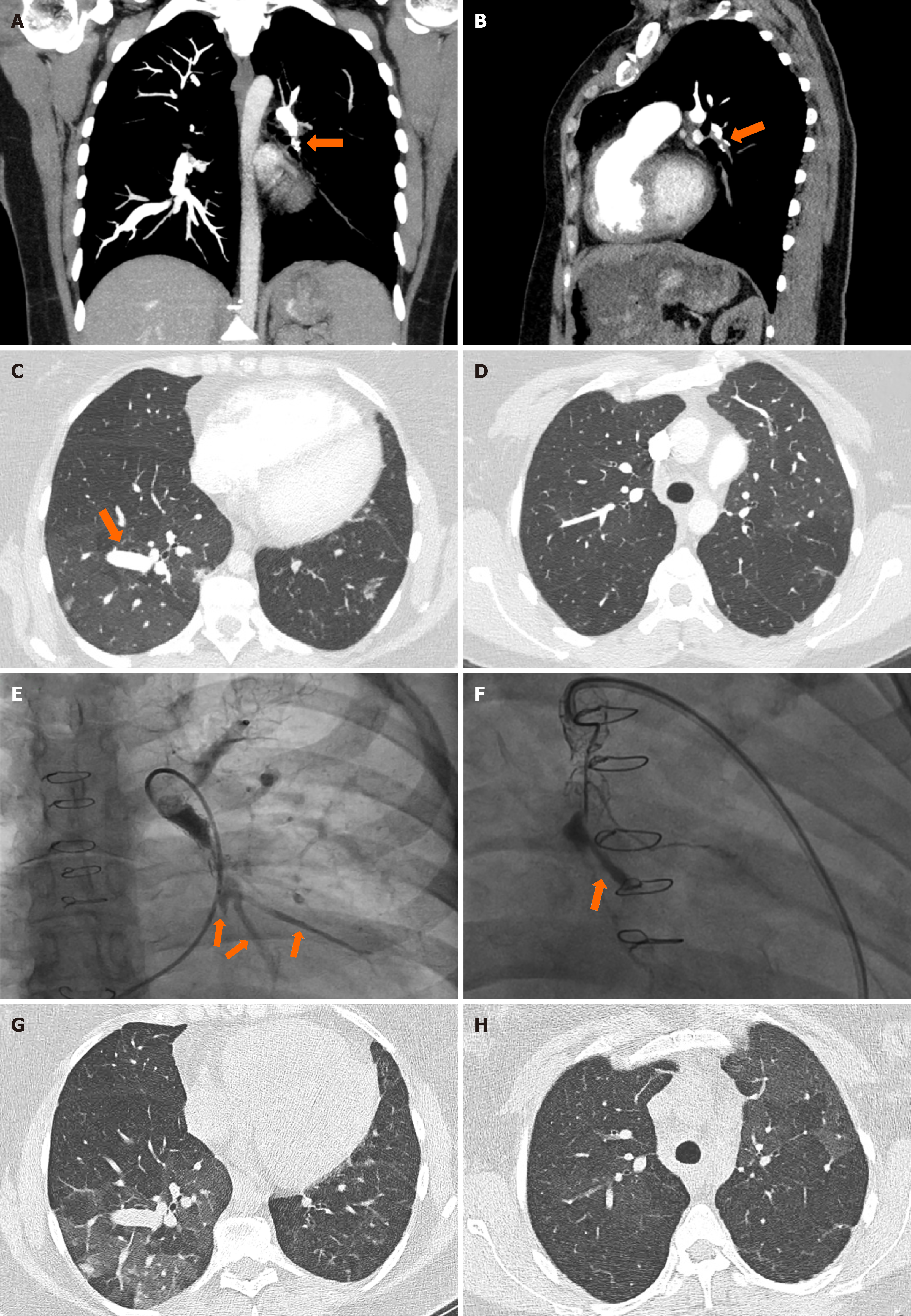
Figure 1 Imaging examinations of the patient.
A: Axial view; B: Sagittal view of a chest computerized tomography (CT) with contrast, mediastinal window, showing lack of propagation of contrast material in the left lower lobe branch of the left pulmonary artery; C: Axial view of a chest CT with contrast, lung window, lower lobe view, demonstrating a widened blood vessel in the right lower lung and ground glass opacities throughout; D: Axial view of a chest CT with contrast, lung window, upper lobe view, demonstrating a widened blood vessel in the right lower lung and mosaicism of lung parenchyma; E: Right heart catheterization (October 2022) showing contrast propagation through 3 arteries distal to the left pulmonary artery stent; F: Right heart catheterization (February 2023) showing complete occlusion of the arteries distal to the fractured and thrombosed stent in the left pulmonary artery; G and H: Axial view of a high-resolution chest CT, November 2022, ground-glass opacities and mosaicism bilaterally.
- Citation: Izhakian S, Korlansky M, Rosengarten D, Bruckheimer E, Kramer MR. Pulmonary artery stent thrombosis and symptomatic pulmonary hypertension following COVID-19 infection in Alagille patient: A case report. World J Clin Cases 2025; 13(9): 96897
- URL: https://www.wjgnet.com/2307-8960/full/v13/i9/96897.htm
- DOI: https://dx.doi.org/10.12998/wjcc.v13.i9.96897


