Copyright
©The Author(s) 2025.
World J Clin Cases. Feb 26, 2025; 13(6): 94330
Published online Feb 26, 2025. doi: 10.12998/wjcc.v13.i6.94330
Published online Feb 26, 2025. doi: 10.12998/wjcc.v13.i6.94330
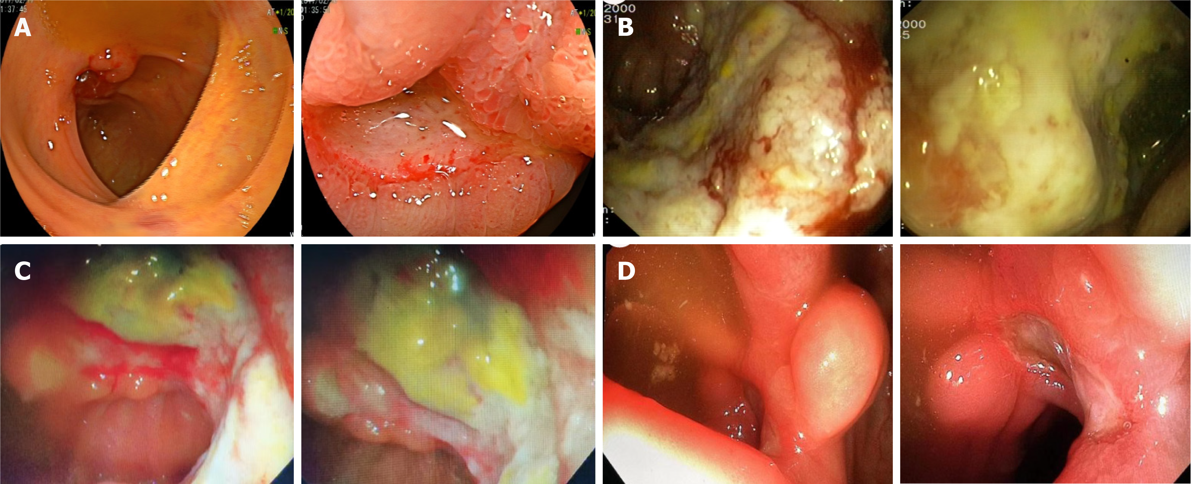
Figure 1 Ileocecal ulcers.
A: The first colonoscopy showing an ileocecal ulcer; B: The second colonoscopy showing a giant irregular ulcer on the ileocecal valve with an ill-defined edge and a dirty coating on the surface; C: The third colonoscopy showing a giant irregular ulcer near the ileocecal valve, measuring approximately 3.0 cm × 5.0 cm, with an ill-defined edge and a dirty coating on the surface; D: The fourth colonoscopy showing an irregular ulcer near the ileocecal valve, measuring approximately 0.6 cm × 1.8 cm, with an ill-defined edge, indicating that the ulcer improved.
- Citation: Yuan WJ, Zheng YJ, Zhang BR, Lin YJ, Li Y, Qiu YY, Yu XP. Hydroxyurea-related ileocecal region ulcers as a rare complication: A case report. World J Clin Cases 2025; 13(6): 94330
- URL: https://www.wjgnet.com/2307-8960/full/v13/i6/94330.htm
- DOI: https://dx.doi.org/10.12998/wjcc.v13.i6.94330


