Copyright
©The Author(s) 2025.
World J Clin Cases. Feb 26, 2025; 13(6): 100673
Published online Feb 26, 2025. doi: 10.12998/wjcc.v13.i6.100673
Published online Feb 26, 2025. doi: 10.12998/wjcc.v13.i6.100673
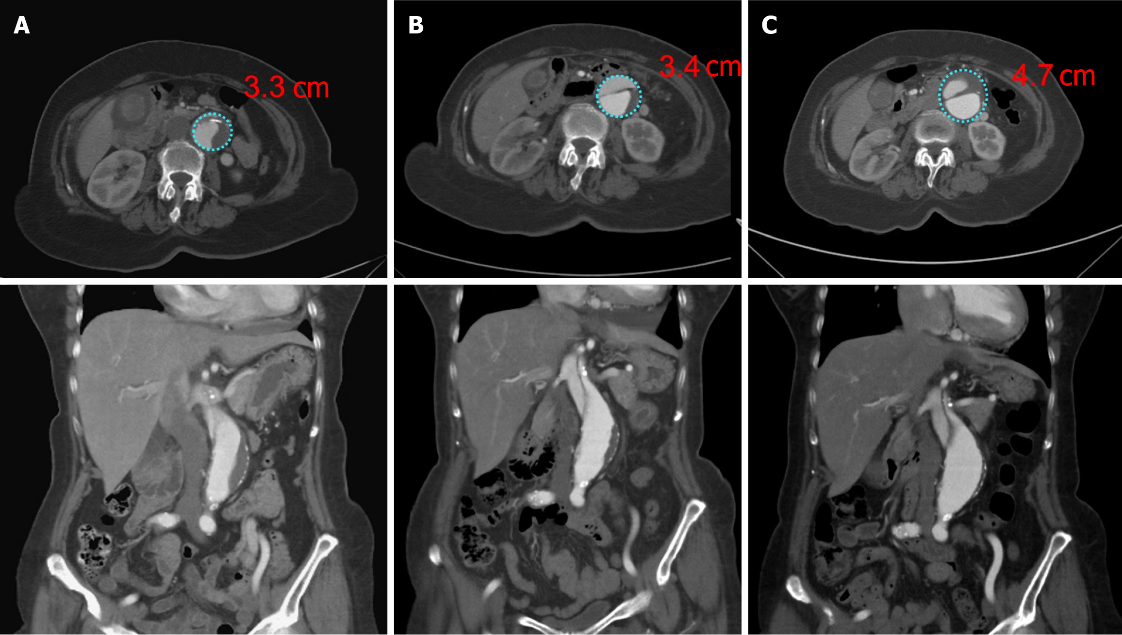
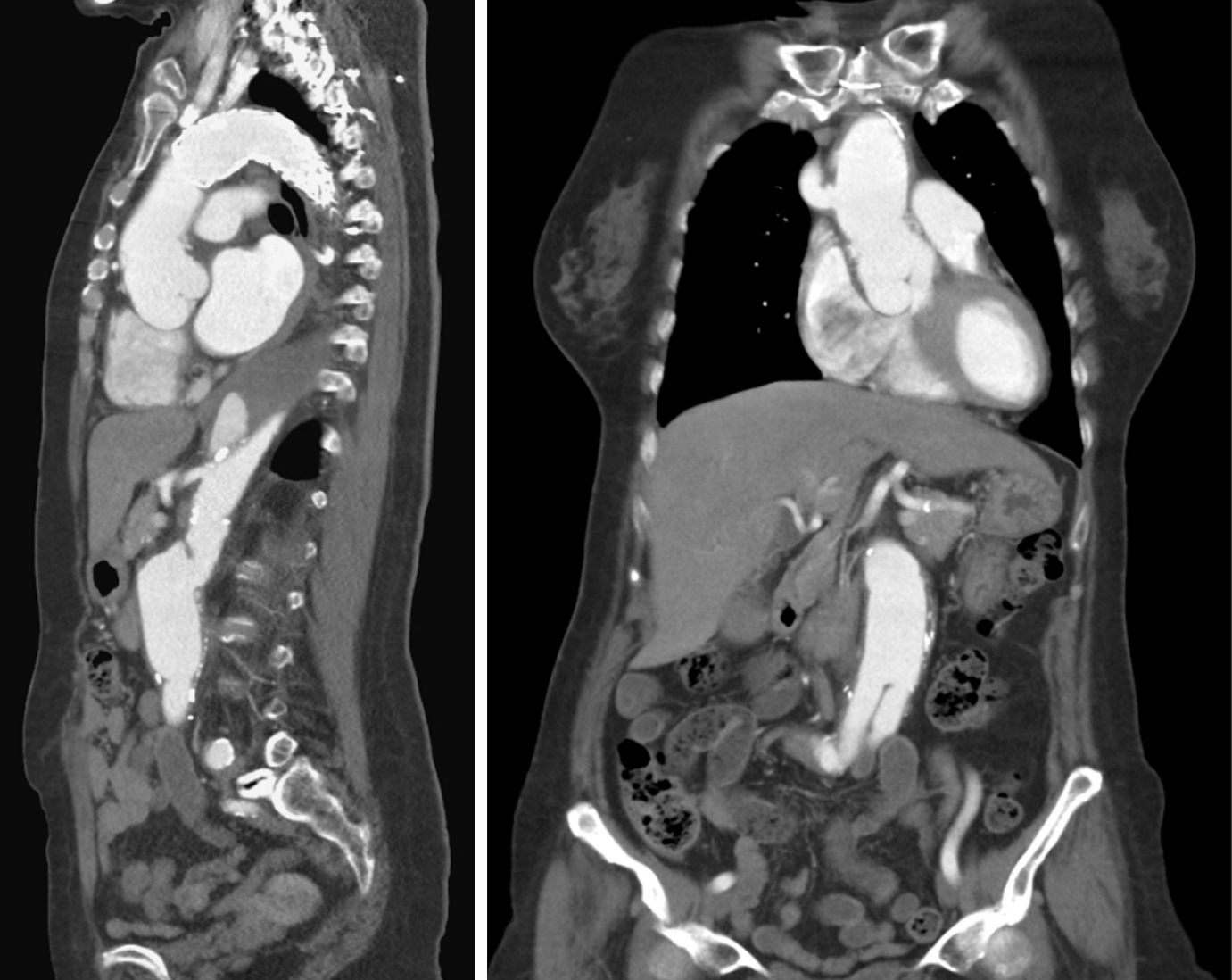
Figure 1 Progression of infra-renal abdominal aortic aneurysm: Sequential computed tomography imaging demonstrates the progression of an infra-renal abdominal aortic aneurysm, which increased in size by over 1 cm within approximately a year.
A: Pre-operative status; B: Post-operative status 2 months later; C: A follow-up 1 year and 4 months post-operation, respectively.
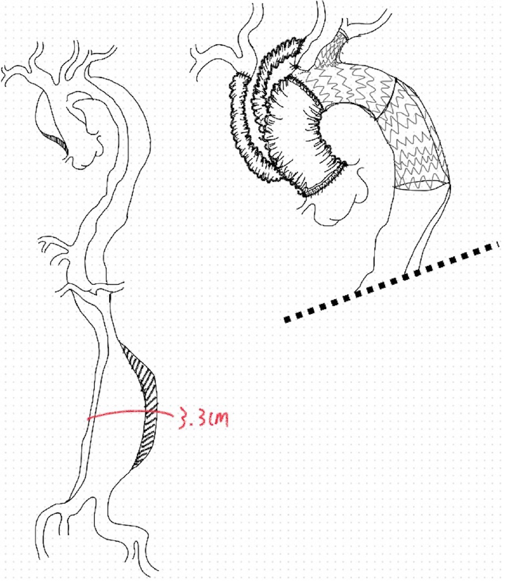
Figure 2 Emergent total arch replacement and thoracic endovascular aortic repair procedure: Involving a total arch replacement using a 28 mm 4-branch Gelsoft™ graft for reconstruction of the ascending aorta and innominate and left carotid arteries.
The procedure included a thoracic endovascular aortic repair with a zone 1 landing using GORE C-TAG devices, fenestration of the left subclavian artery with a VIABAHN stent, and resuspension of the aortic valve. An aortic aneurysm measuring approximately 3.3 cm was noted in the abdominal aorta at the time of operation.
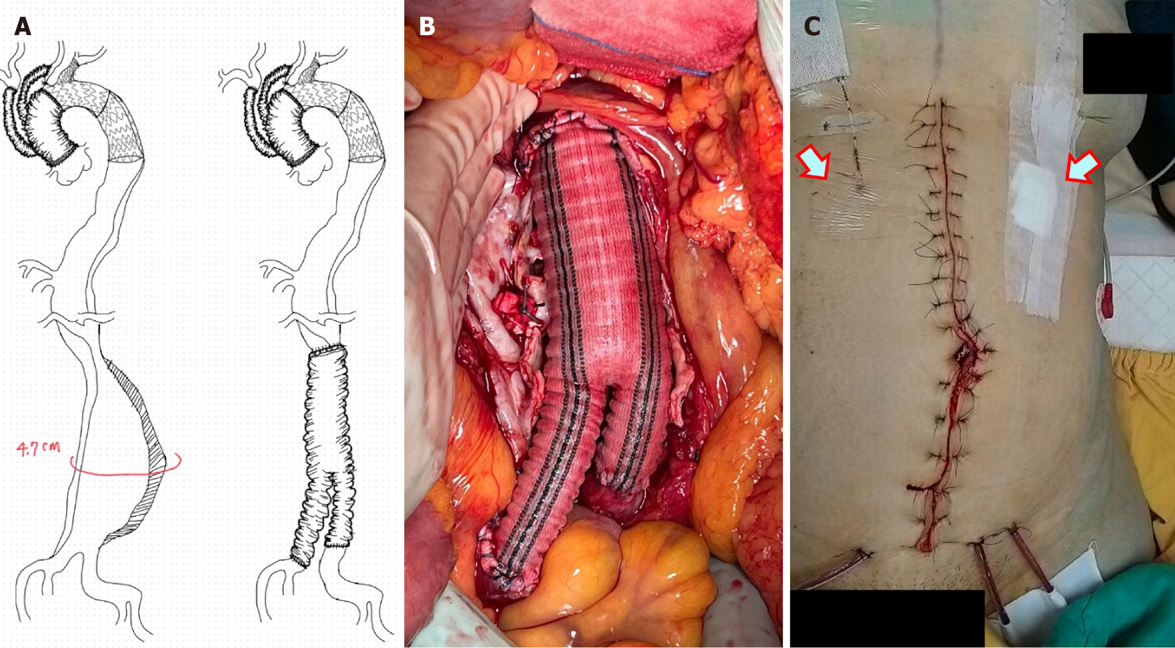
Figure 3 Second operation utilizing Gelsoft™ vascular prosthesis: Surgical intervention involved the use of a Gelsoft™ vascular pro
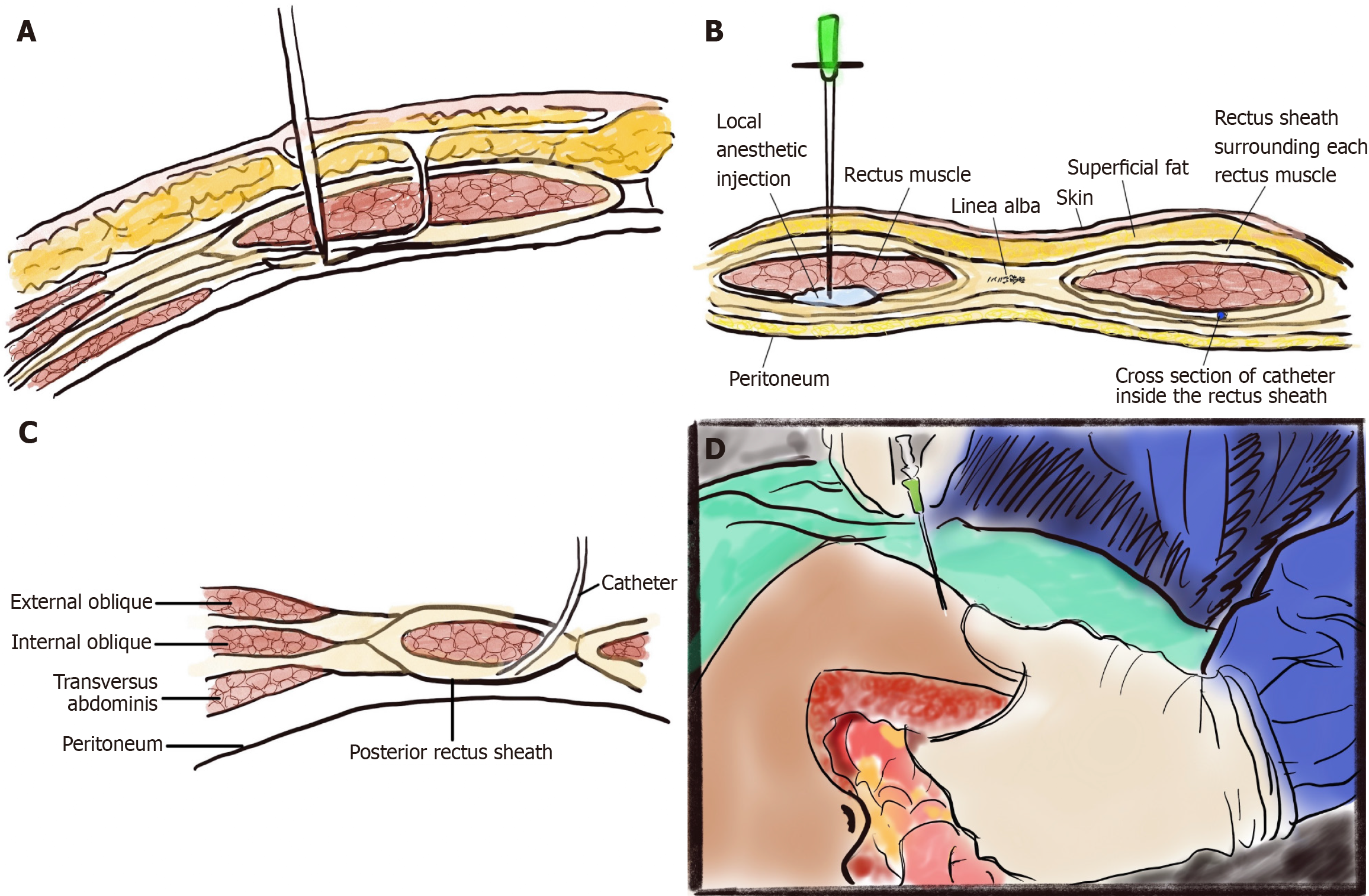
Figure 4 Rectus sheath block technique: A detailed cross-sectional diagram of the abdominal wall illustrating the technique for rectus sheath block.
A: The needle target; B: The transverse section of the anterior abdominal wall depicting needle position and local anesthetic injection; C: The placement of the rectus sheath catheter; D: The catheter-over-needle assembly being inserted in a cephalad-to-caudad direction, with intra-abdominal hand placement to feel for needle advancement.
Figure 5 Follow-up computed tomography scans showing stable condition.
This image confirms the absence of progression or complications related to the previously observed abdominal aortic aneurysm and surgical interventions.
- Citation: Chen KH, Kang MY, Chang YT, Huang SY, Wu YS. Enhancing postoperative pain control by surgically-initiated rectus sheath block in abdominal aortic aneurysm open repair: A case report. World J Clin Cases 2025; 13(6): 100673
- URL: https://www.wjgnet.com/2307-8960/full/v13/i6/100673.htm
- DOI: https://dx.doi.org/10.12998/wjcc.v13.i6.100673


