Copyright
©The Author(s) 2025.
World J Clin Cases. Feb 16, 2025; 13(5): 99963
Published online Feb 16, 2025. doi: 10.12998/wjcc.v13.i5.99963
Published online Feb 16, 2025. doi: 10.12998/wjcc.v13.i5.99963
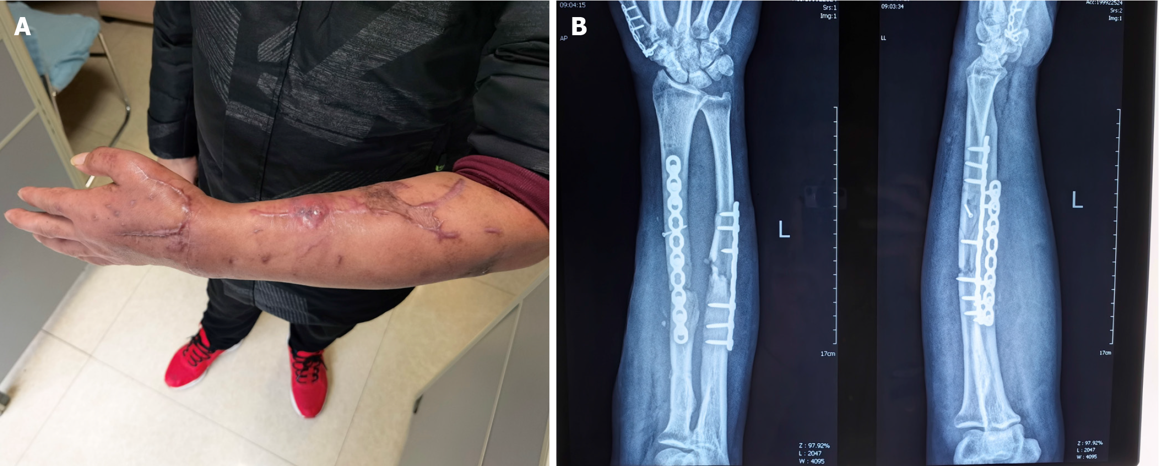
Figure 1 Presentation of the patient.
A: Skin sinus of the patient; B: Preoperative X-ray showing the infectious nonunion of the left radius.
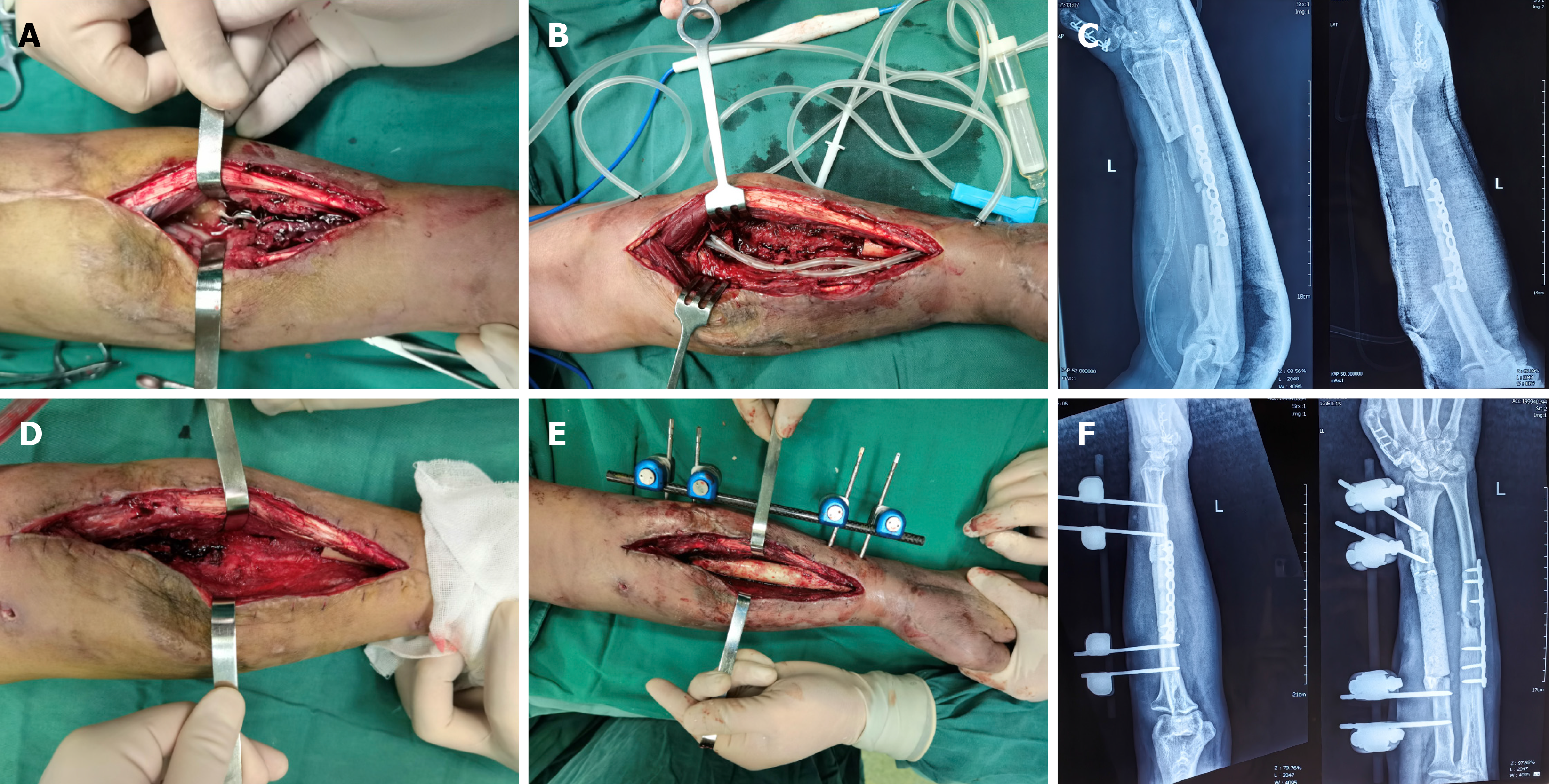
Figure 2 Debridement and cement implantation to the patient.
A: Infectious focus of the left radius; B: Removal of the implant and infectious bone segment; and placement of drainage tubes and vacuum sealing drainage; C: X-ray after debridement; D: Effective resolution of the focus; E: Placement of the antibiotic spacer and external fixation; F: X-ray after placement of the antibiotic spacer and external fixation.
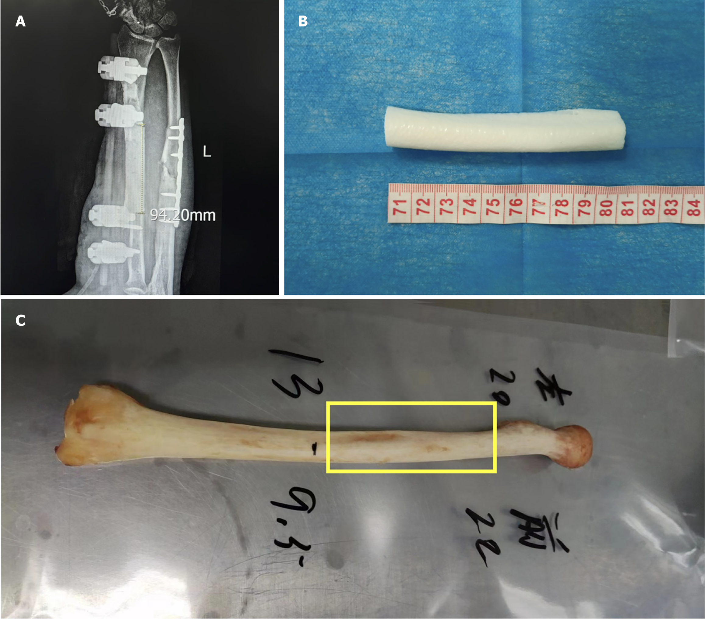
Figure 3 Bone defect and selection of the donor bone segment.
A: Length of the left radius defect; B: 3D-printed model of the defective bone segment; C: Donor bone graft matched to the defective radius.
Figure 4 The induced membrane and X-ray after bone implantation.
A: Cavity and induced membrane after spacer removal; B: Postoperative X-ray; C: Length of the allogeneic cortical bone graft.
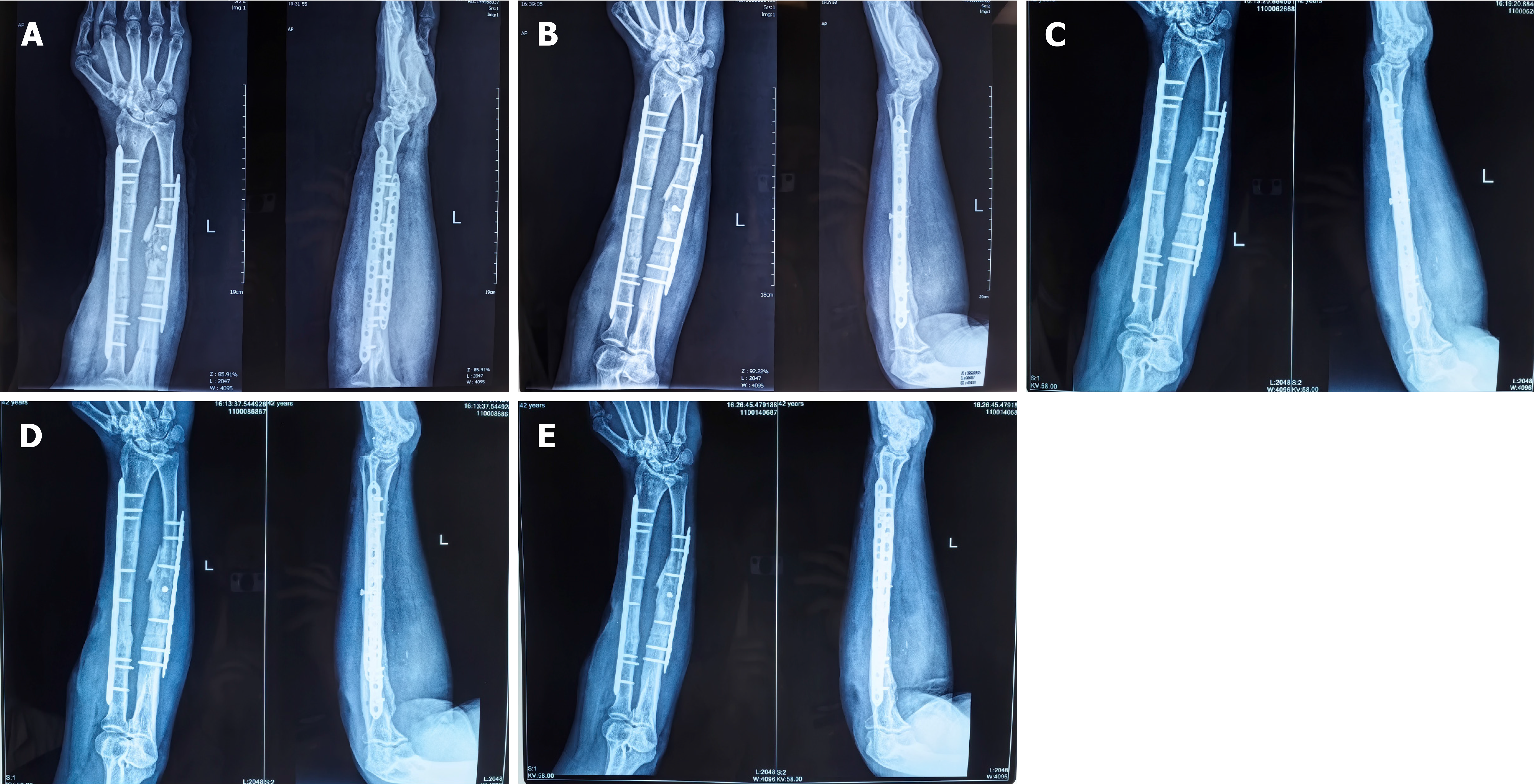
Figure 5 Radiographs at follow-up.
A: X-ray image after 3 months; B: X-ray image after 6 months; C: X-ray image after 10 months; D: X-ray image after 14 months; E: X-ray image after 24 months.
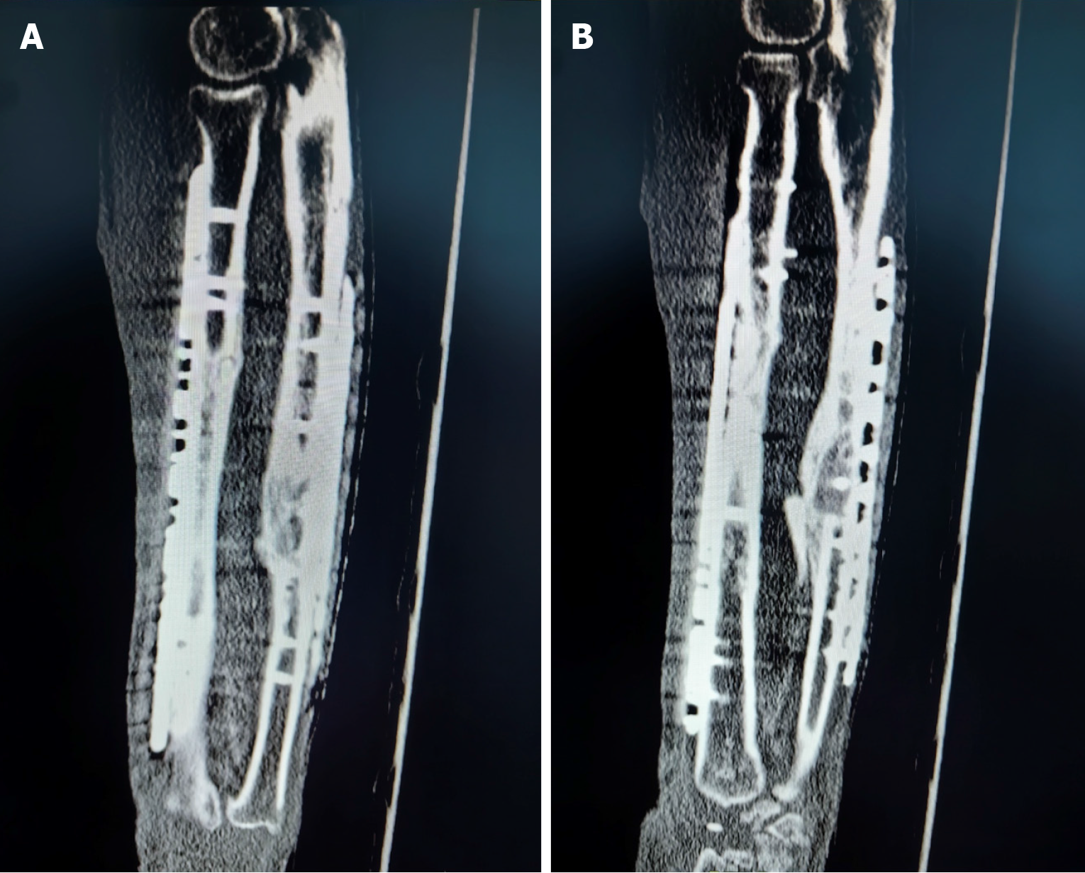
Figure 6 Healing of the bone graft.
A: Computed tomography (CT) scan after 14 months, demonstrating proximal healing of the allogenic cortical bone graft; B: CT scan after 14 months, demonstrating distal healing of the allogenic cortical bone graft.
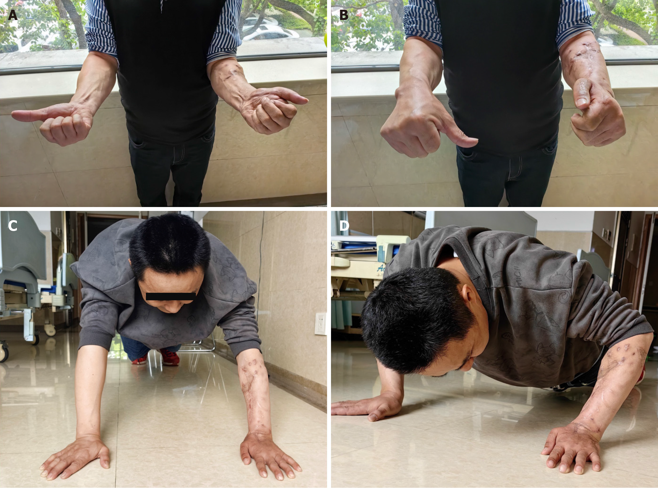
Figure 7 Functional recovery of the patient at 2 years follow-up.
A: Supination function of the forearm; B: pronation function of the forearm; C: The support function of the forearm; D: Strength of the forearm.
- Citation: Zong HY, Liu Y, Yin X, Zhou W, Li N. Masquelet technique using an allogeneic cortical bone graft for a large bone defect: A case report. World J Clin Cases 2025; 13(5): 99963
- URL: https://www.wjgnet.com/2307-8960/full/v13/i5/99963.htm
- DOI: https://dx.doi.org/10.12998/wjcc.v13.i5.99963


