Copyright
©The Author(s) 2025.
World J Clin Cases. Oct 16, 2025; 13(29): 110454
Published online Oct 16, 2025. doi: 10.12998/wjcc.v13.i29.110454
Published online Oct 16, 2025. doi: 10.12998/wjcc.v13.i29.110454
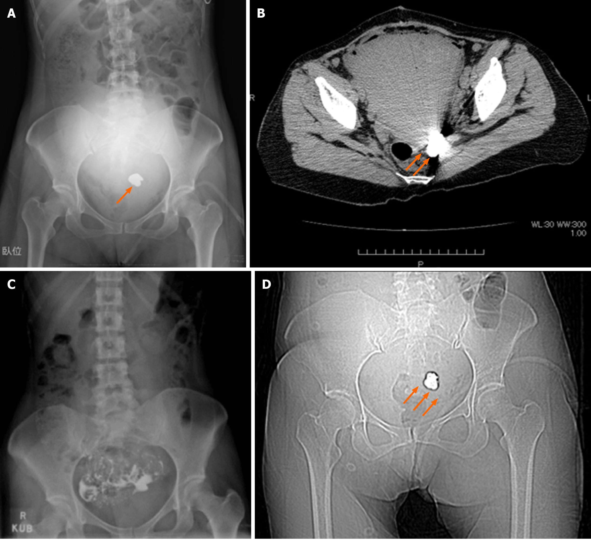
Figure 1 Imaging of this case.
A: Abdominal radiograph taken immediately after cesarean section. A near-round, homogeneously hyperdense area with a regular margin can be seen in the pelvic cavity, with radiolucency similar to that of metals (as indicated by the arrow); B: Normal abdominal computed tomography scan (taken immediately after surgery). The computed tomography value of the mass-like shadow in the pelvic cavity was 7000 Hounsfield units, which is similar to that of metals (indicated by the double arrow); C: Hysterosalpingogram performed previously at another facility using an oil-based iodinated contrast agent; D: Plain abdominal radiograph. The mass-like shadow in the pelvic cavity appears deformed and reduced in size (indicated by the triple arrow). Hysterosalpingogram using an oil-based iodinated contrast medium was performed at another facility previously (sly at another facility).
- Citation: Morita A, Kakinuma T, Segawa A, Harada S, Takae S, Tamura M, Suzuki N. Prolonged retention of oil-based iodinated contrast medium observed on plain abdominal radiograph after cesarean section: A case report. World J Clin Cases 2025; 13(29): 110454
- URL: https://www.wjgnet.com/2307-8960/full/v13/i29/110454.htm
- DOI: https://dx.doi.org/10.12998/wjcc.v13.i29.110454


