Copyright
©The Author(s) 2025.
World J Clin Cases. Sep 16, 2025; 13(26): 108308
Published online Sep 16, 2025. doi: 10.12998/wjcc.v13.i26.108308
Published online Sep 16, 2025. doi: 10.12998/wjcc.v13.i26.108308
Figure 1 Three-dimensional visualization reconstruction.
A: Three-dimensional reconstructed image; B: Cross section of the abdominal computed tomography (CT); C: Coronal view of the abdominal CT; D: Sagittal view of the abdominal CT.
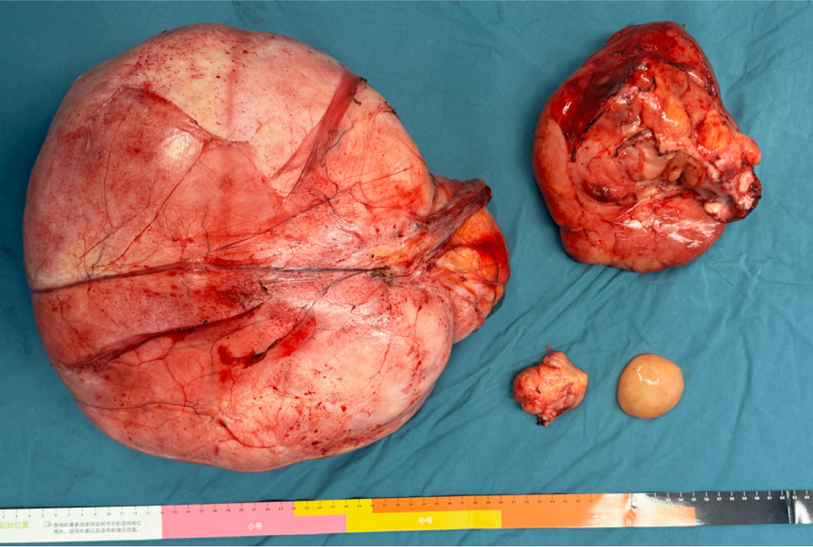
Figure 2
Postoperative tissue specimen (maximum 35 cm × 28 cm × 14 cm).
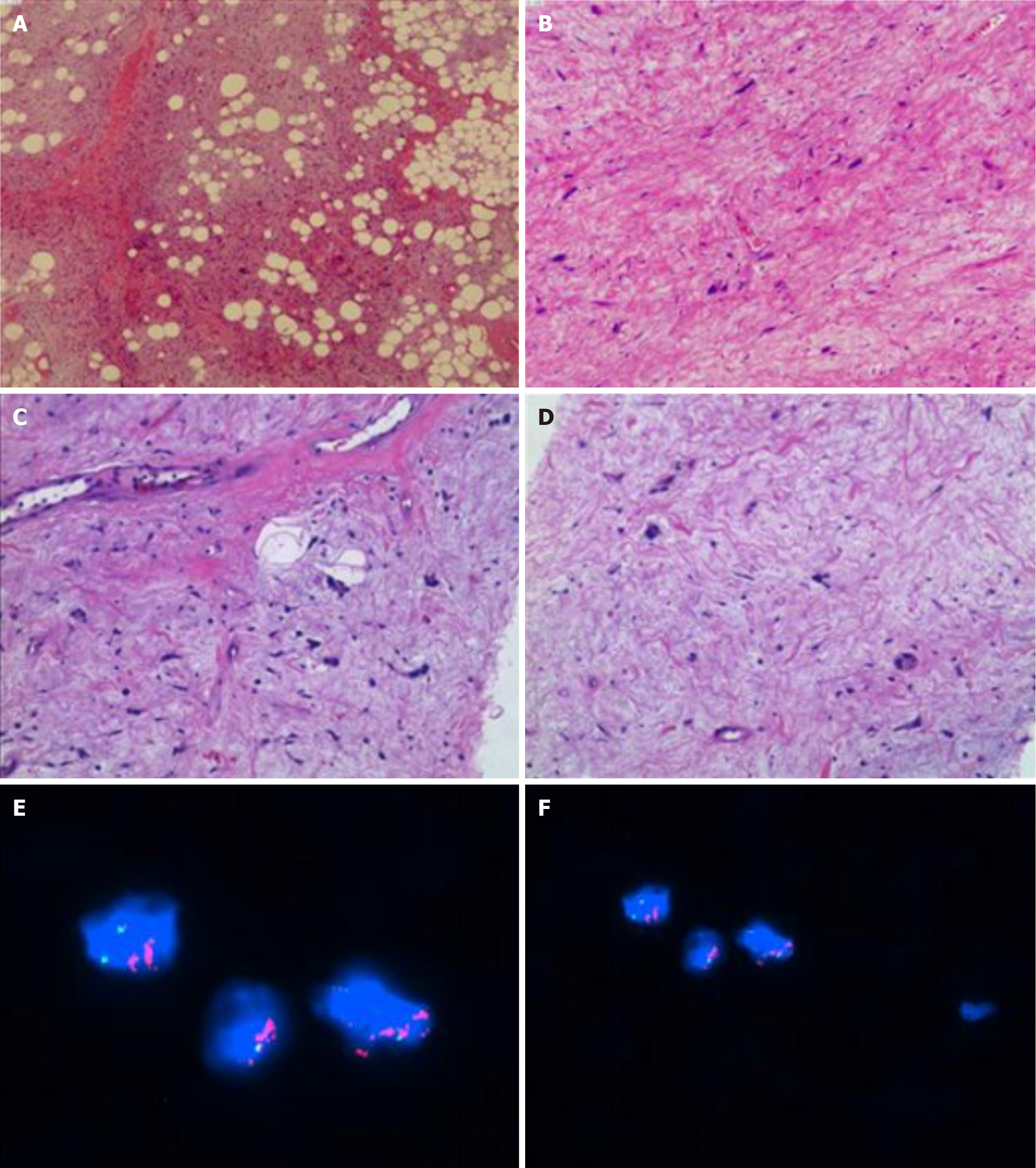
Figure 3 Molecular detection revealed mouse double minute 2 gene expansion.
A and B: Tissue staining (hematoxylin and eosin 10 × 20); C and D: Immunohistochemistry; E and F: Molecular detection.
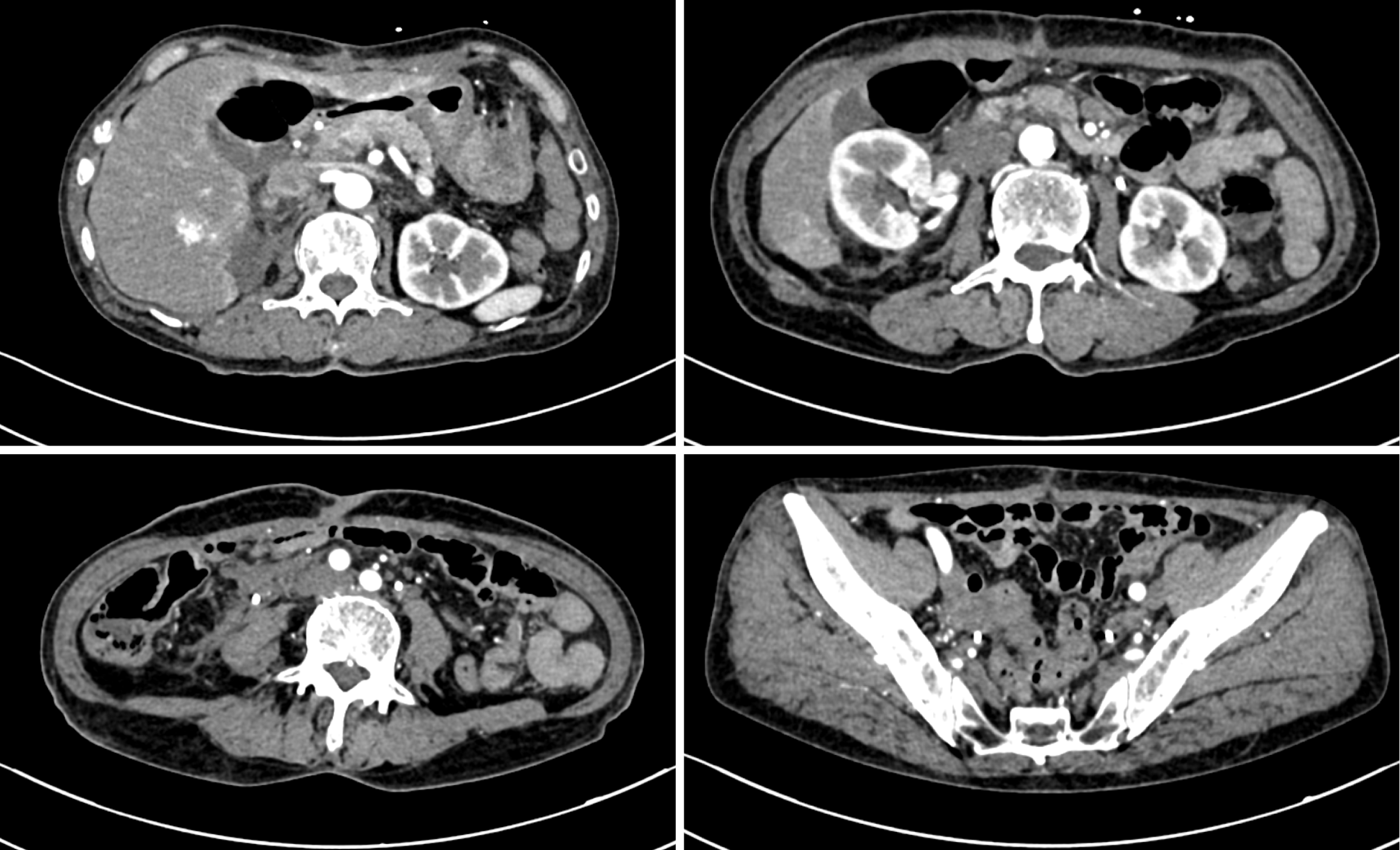
Figure 4
Contrast-enhanced abdominal computed tomography after 2 months.
- Citation: Cheng LL, Tang B, Liu H, Zhu F, Chen YF, Zhang W. Thoughts and challenges of giant retroperitoneal liposarcoma: A case report. World J Clin Cases 2025; 13(26): 108308
- URL: https://www.wjgnet.com/2307-8960/full/v13/i26/108308.htm
- DOI: https://dx.doi.org/10.12998/wjcc.v13.i26.108308


