Copyright
©The Author(s) 2025.
World J Clin Cases. Sep 16, 2025; 13(26): 105486
Published online Sep 16, 2025. doi: 10.12998/wjcc.v13.i26.105486
Published online Sep 16, 2025. doi: 10.12998/wjcc.v13.i26.105486
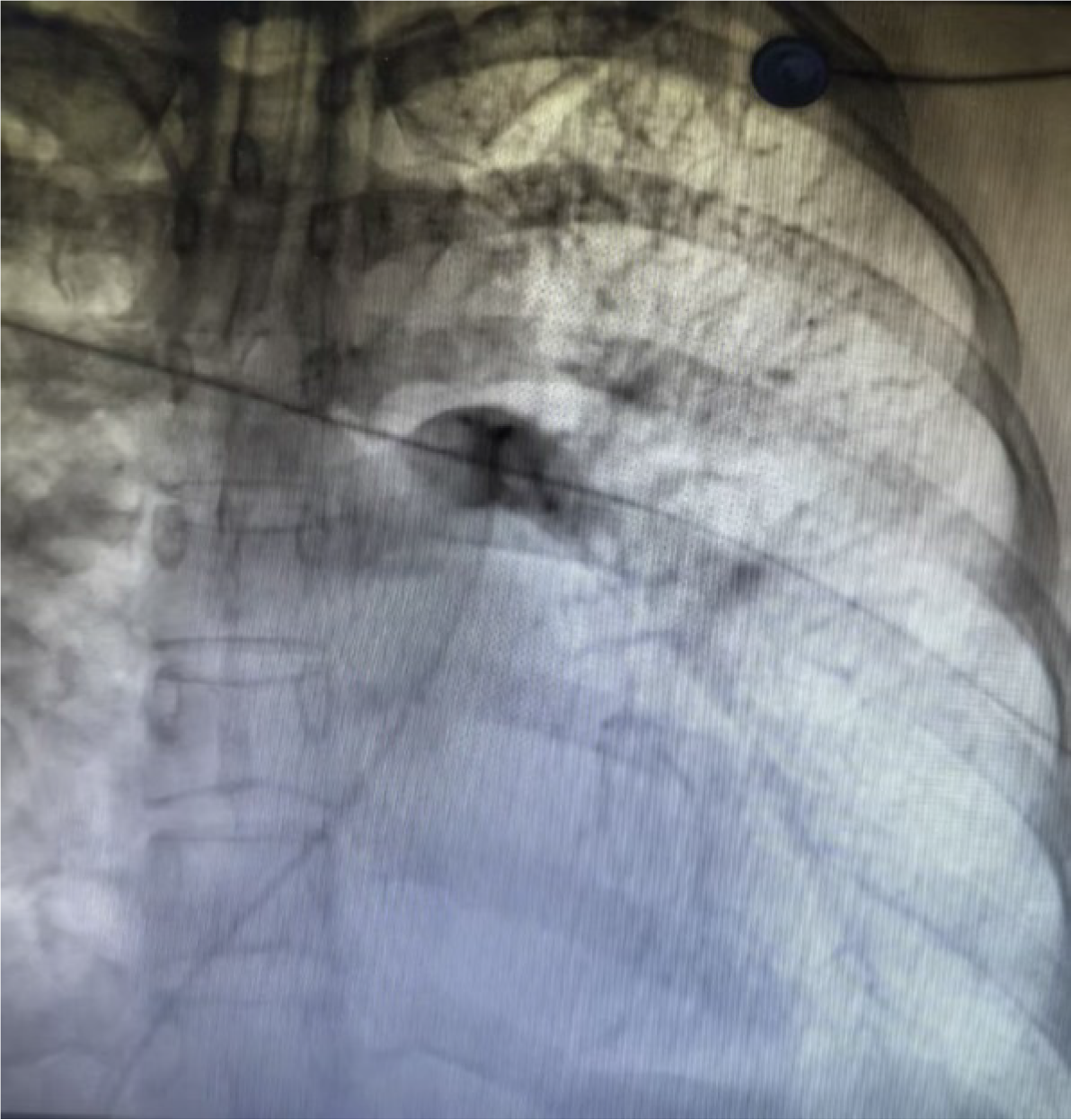
Figure 1
The main pulmonary artery and its distal branches were not shown by angiography.
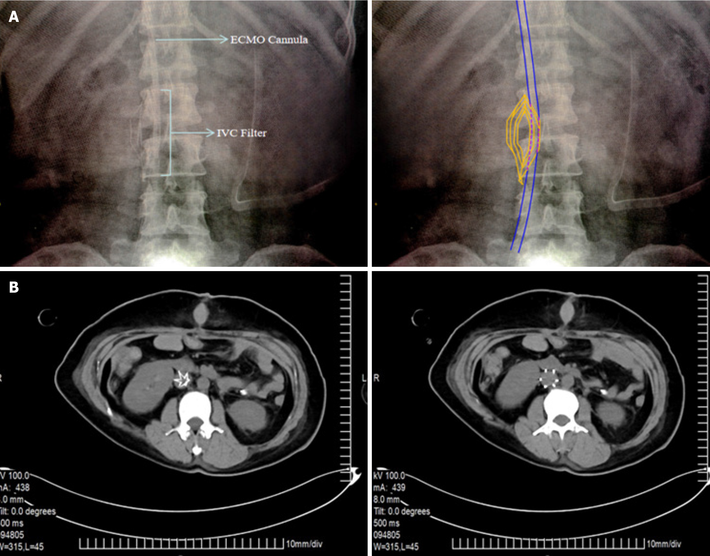
Figure 2 Imaging findings.
A: The extracorporeal membrane oxygenation venous drainage catheter travels in the inferior vena cava through the hollow region of the filter, which is located at the L2 level; B: After extracorporeal membrane oxygenation weaning and removal of the venous drainage catheter, the inferior vena cava filter remained at the L2 level with no thrombus inside or outside the filter.
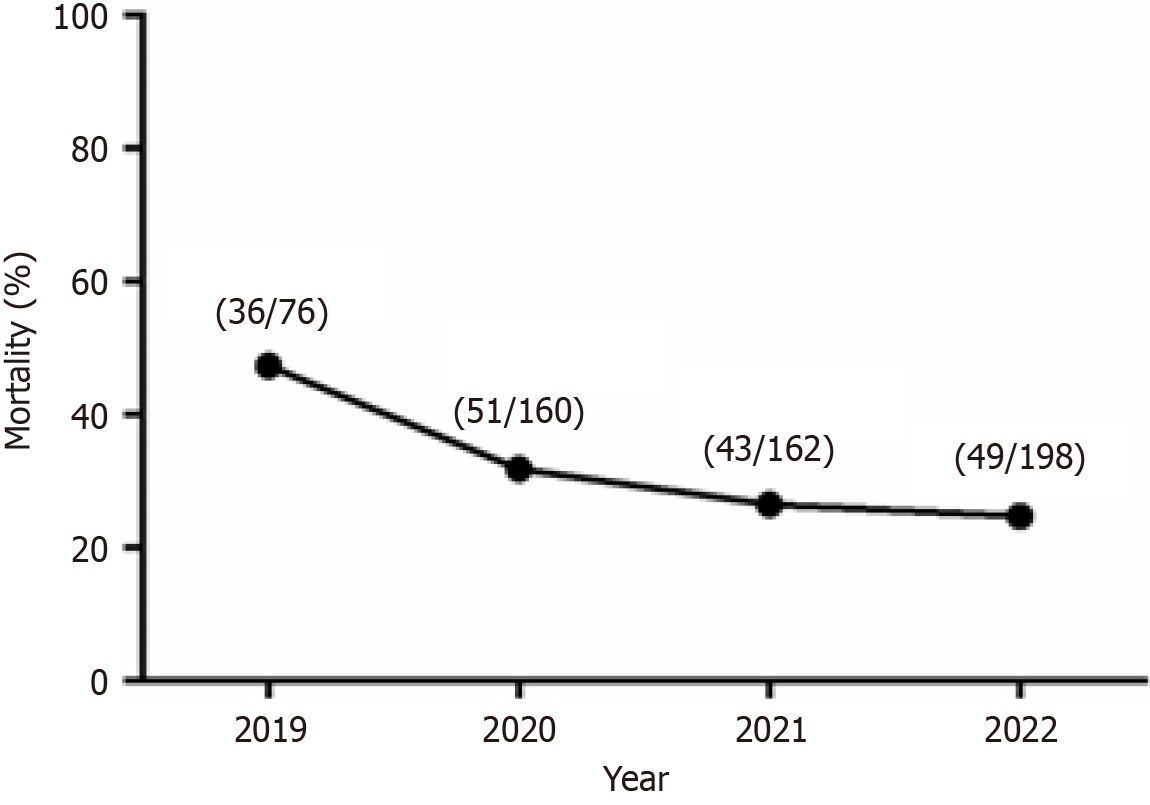
Figure 3 Mortality of patients diagnosed with pulmonary thromboembolism in the Affiliated Hospital of Zunyi Medical University from 2019 to 2022 (n1/n2).
n1 represents the annual number of deaths and n2 represents the annual number of confirmed pulmonary thromboembolism cases.
- Citation: Gao F, Ma S, Xiao X, Yang H, Qian MJ. Emergency veno-arterial extracorporeal membrane oxygenation cannulation through the femoral vein with a pre-positioned inferior vena cava filter: A case report. World J Clin Cases 2025; 13(26): 105486
- URL: https://www.wjgnet.com/2307-8960/full/v13/i26/105486.htm
- DOI: https://dx.doi.org/10.12998/wjcc.v13.i26.105486


