Copyright
©The Author(s) 2025.
World J Clin Cases. Aug 6, 2025; 13(22): 106122
Published online Aug 6, 2025. doi: 10.12998/wjcc.v13.i22.106122
Published online Aug 6, 2025. doi: 10.12998/wjcc.v13.i22.106122
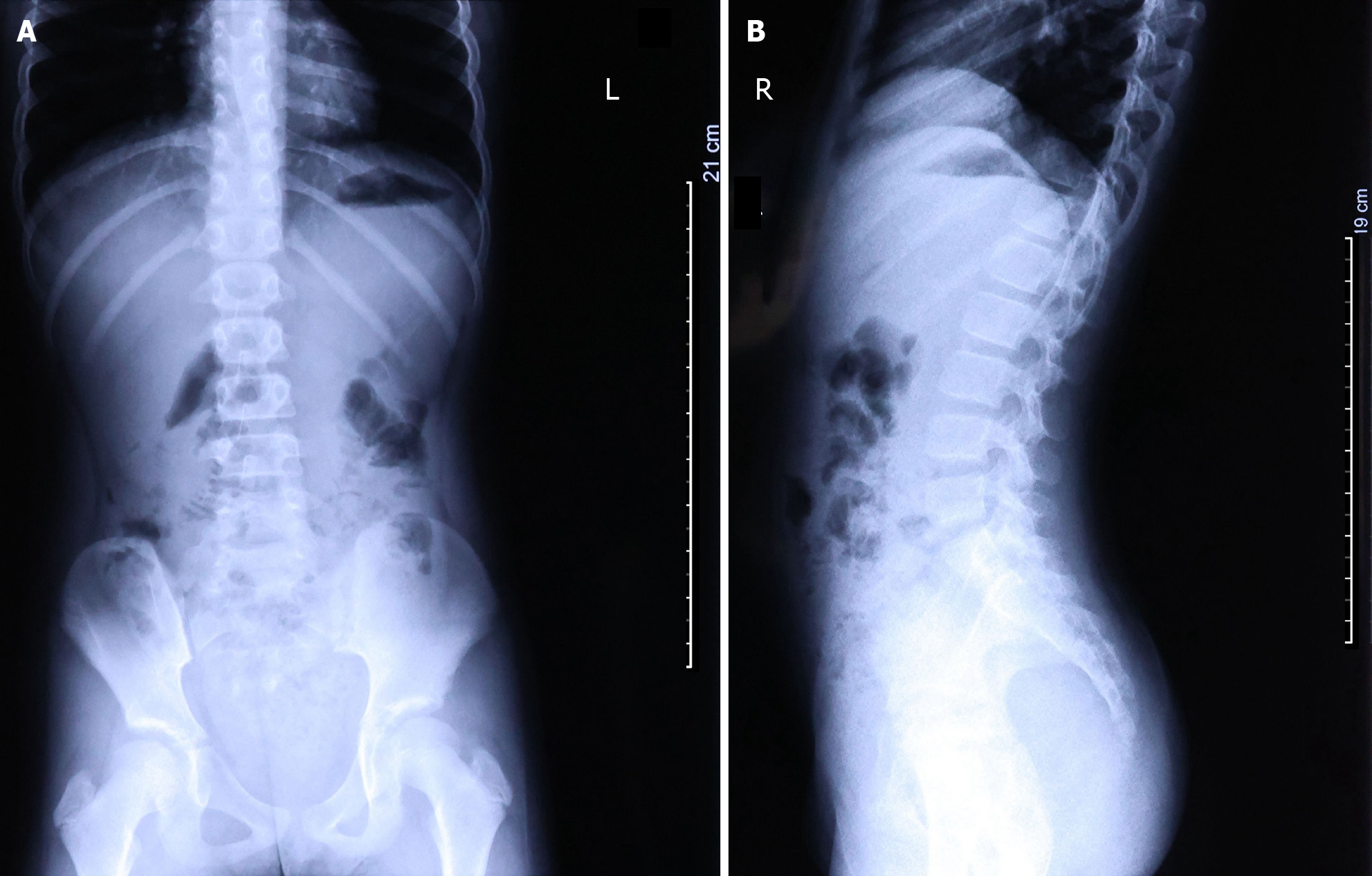
Figure 1 X-ray examination revealed poor aeration of the small intestine in the lower abdomen, along with the presence of air-fluid levels.
A: Front view; B: Lateral view.
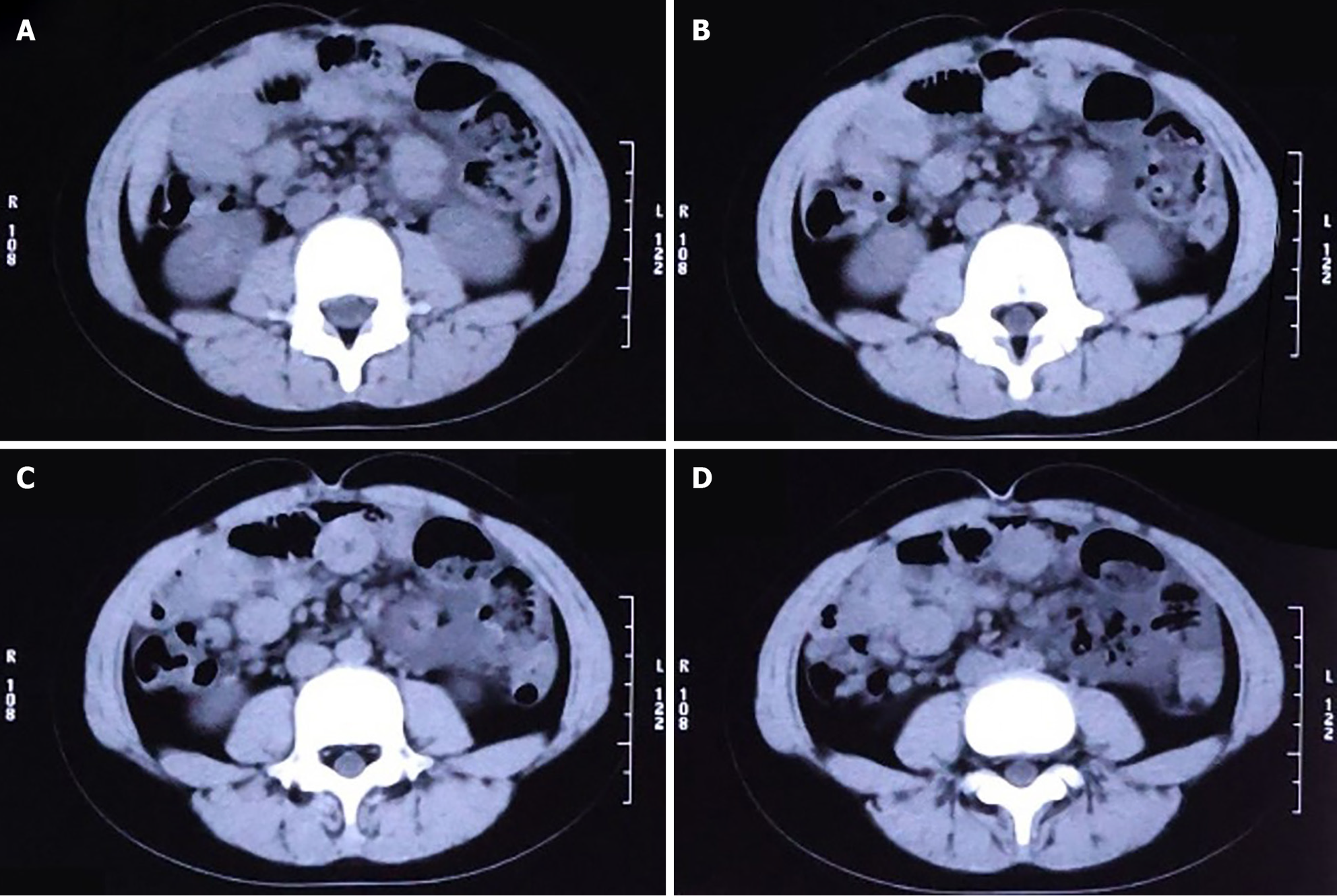
Figure 2 Computed tomography scan reveals a definitive obstruction site, along with visible exudation around the intestinal tract.
A: The intestinal tract is disorganized and air-fluid levels are also observed; B: The obstruction point is located below and to the left of the umbilicus; C: The intestinal wall at the point of obstruction is thickened, with pronounced exudation from the surrounding intestinal tract; D: The colon and rectum are collapsed.
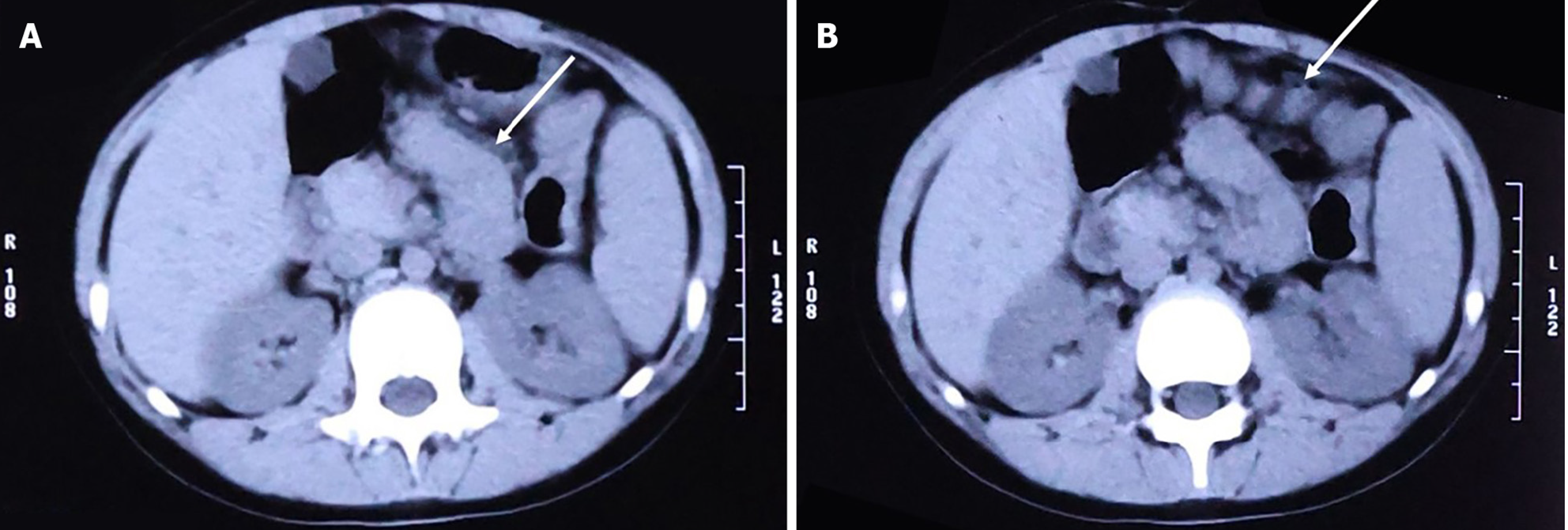
Figure 3 Computed tomography scan reveals soft tissue structures surrounding the small intestine.
A: Soft tissue structures surround the small intestine; B: Small intestine is pulled, exhibiting a distinctive "accordion-like" appearance.
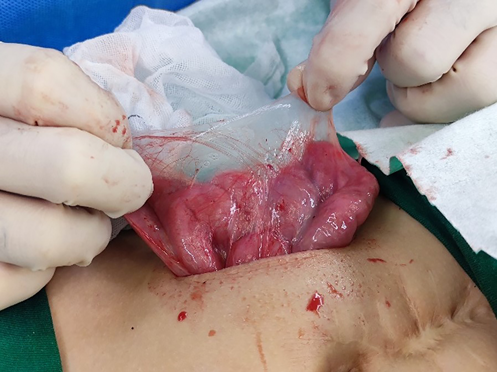
Figure 4 Intraoperative images reveal a substantial membranous structure enveloping the small intestine, with no adhesions observed between the membranous structure and the peritoneum.
- Citation: Zheng HJ, Zhang JD, Wang ZC, Yao LY. Abdominal cocoon syndrome in a 10-year-old young adolescent after abdominal operation: A case report and review of literature. World J Clin Cases 2025; 13(22): 106122
- URL: https://www.wjgnet.com/2307-8960/full/v13/i22/106122.htm
- DOI: https://dx.doi.org/10.12998/wjcc.v13.i22.106122


