Copyright
©The Author(s) 2025.
World J Clin Cases. Aug 6, 2025; 13(22): 104924
Published online Aug 6, 2025. doi: 10.12998/wjcc.v13.i22.104924
Published online Aug 6, 2025. doi: 10.12998/wjcc.v13.i22.104924
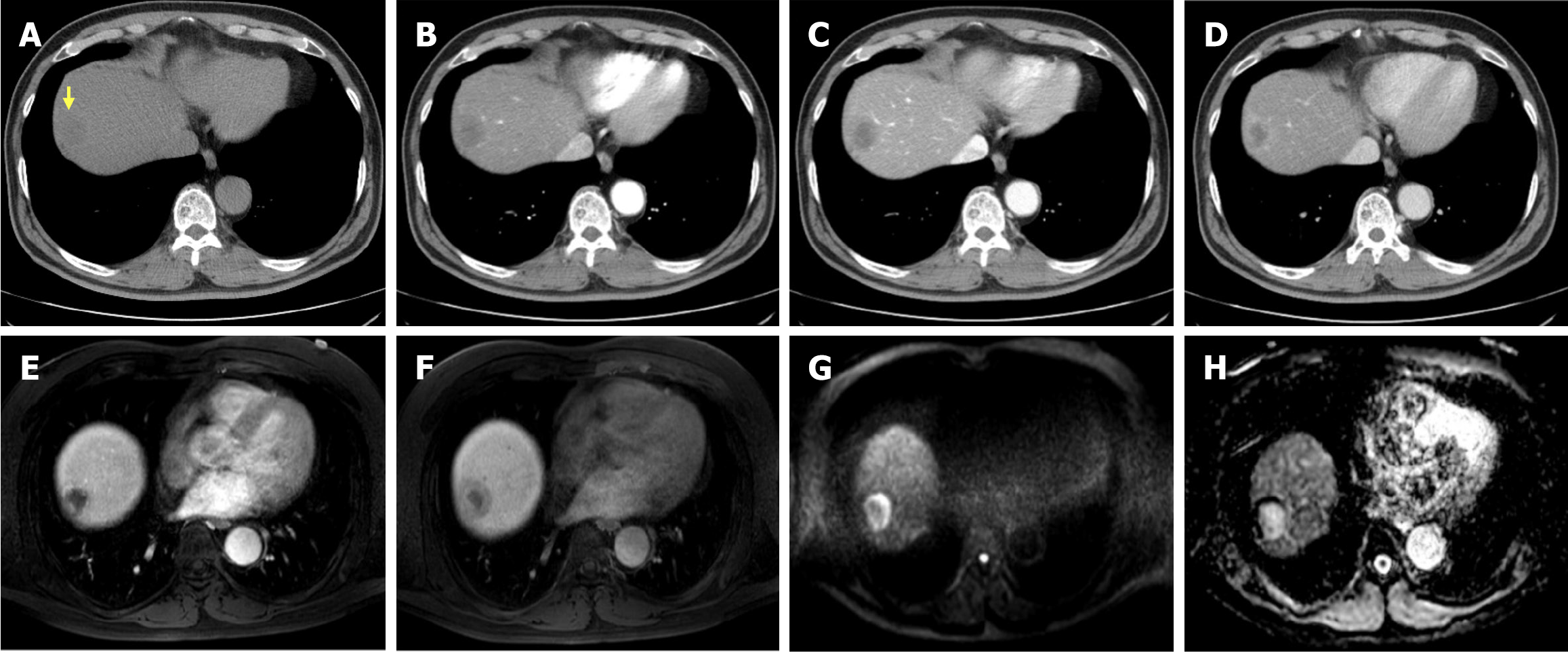
Figure 1 Preoperative abdominal dynamic computed tomography and liver magnetic resonance imaging findings.
A and B: A low-attenuation nodule (yellow arrow) in the right hepatic dome subcapsular area was observed on pre-contrast imaging, showing peripheral rim enhancement in the arterial phase (B). C and D: In the portal phase (C), the nodule demonstrates progressive centripetal enhancement, and the delayed phase (D) shows a typical centripetal enhancement pattern. E and F: Axial T1-weighted arterial-(E) and hepatobiliary-phase (F) images show peripheral enhancement with gradual centripetal filling of contrast media. G and H: Diffusion-weighted imaging with a b-value of 1000 (G) demonstrates marked peripheral high signal intensity, while the apparent diffusion coefficient map (H) reveals high signal intensity in the core of the lesion.
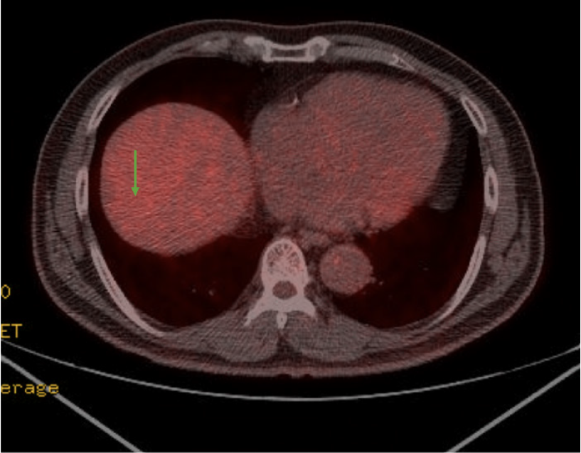
Figure 2 Preoperative positron emission tomography computed tomography findings.
An isometabolic mass in the hepatic dome (green arrow), showing metabolic activity similar to that of the liver parenchyma (SUVmax, 2.7; SUVmean, 2.1), was more suggestive of a benign mass than a malignancy, with no metabolic evidence of local recurrence or regional lymph node involvement.
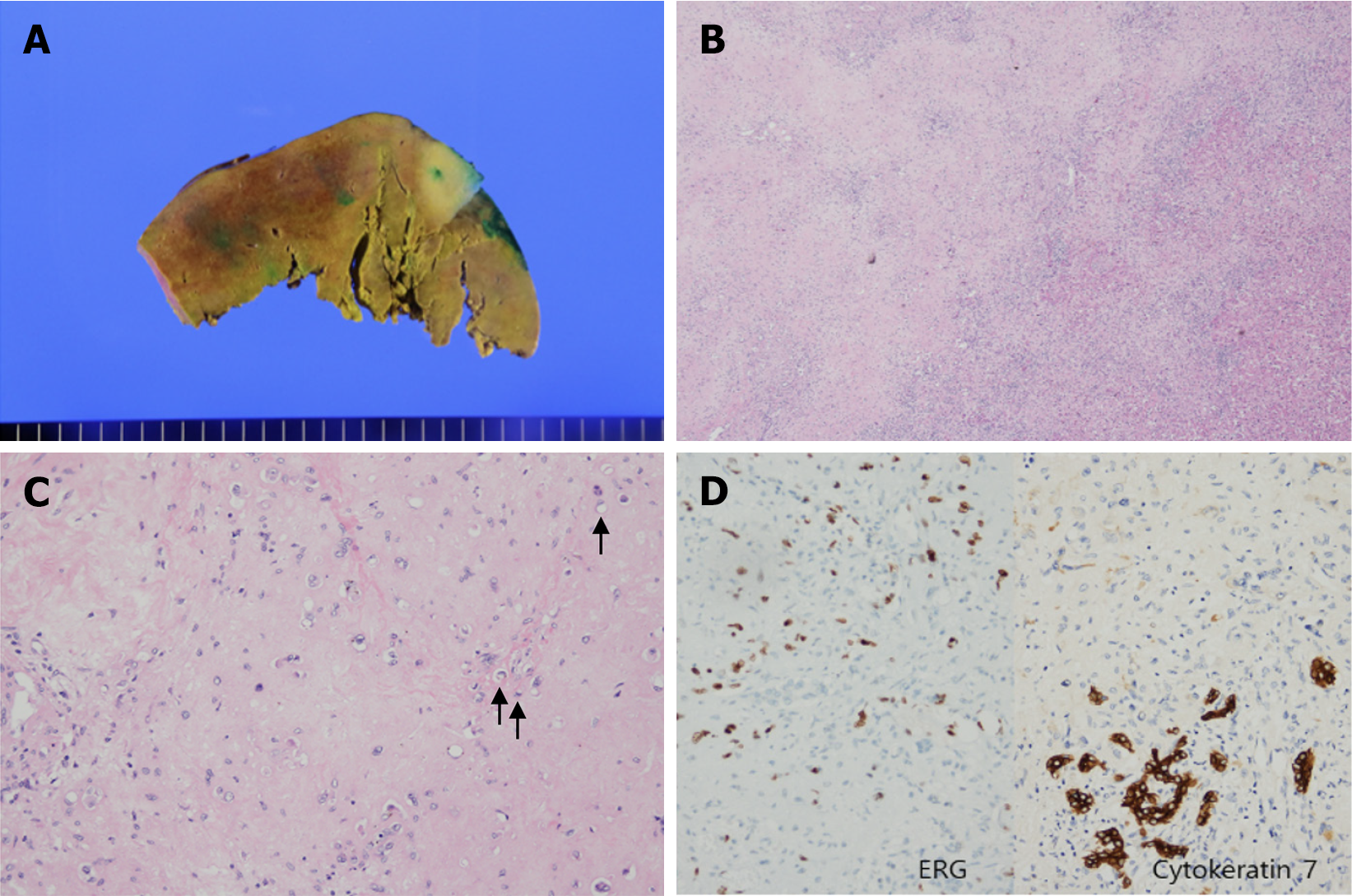
Figure 3 Pathologic findings of hepatic epithelioid hemangioendothelioma.
A: The cut surface shows an ill-defined whitish mass; B: The tumor demonstrates hypercellularity with margins indistinct from the surrounding tissue (H&E stain, × 20); C: Tumor cells are arranged individually or in cords within a myxohyaline stroma, with frequent intracytoplasmic vacuoles (black arrows) (H&E stain, ×200); D: Immunohistochemistry reveals strong reactivity for ERG, indicating a vascular endothelial origin, and also shows immunoreactivity for Cytokeratin 7. The CK7-positive staining pattern appeared morphologically and resembled the intrahepatic bile ducts.
Figure 4 Follow-up abdominal dynamic computed tomography on postoperative day 5.
A: Arterial phase view; B: Hepatobiliary phase axial view; C: Hepatobiliary phase coronal view. These findings demonstrate evidence of laparoscopic anterior resection with a small fluid collection at the resection margin, consistent with the usual postoperative changes.
Figure 5 Follow-up abdominal dynamic computed tomography at 3 years post-surgery.
A: Arterial phase view; B: Hepatobiliary phase axial view; C: Hepatobiliary phase coronal view. There was no evidence of recurrence. The liver showed no newly developed lesions and the patient remained disease-free.
- Citation: Shin SH, Koh YS, Song S. Hepatic epithelioid hemangioendothelioma managed with minimally invasive surgery: A case report. World J Clin Cases 2025; 13(22): 104924
- URL: https://www.wjgnet.com/2307-8960/full/v13/i22/104924.htm
- DOI: https://dx.doi.org/10.12998/wjcc.v13.i22.104924


