Copyright
©The Author(s) 2025.
World J Clin Cases. Aug 6, 2025; 13(22): 104643
Published online Aug 6, 2025. doi: 10.12998/wjcc.v13.i22.104643
Published online Aug 6, 2025. doi: 10.12998/wjcc.v13.i22.104643
Figure 1 Computed tomography.
A: Pelvic Computed tomography with IV contrast revealed a slightly enlarged cervix with the diameter of 3.3 cm, uneven reinforcement area with blurred margins; B and C: Metastasis to distant organs or lymph nodes was not detected.
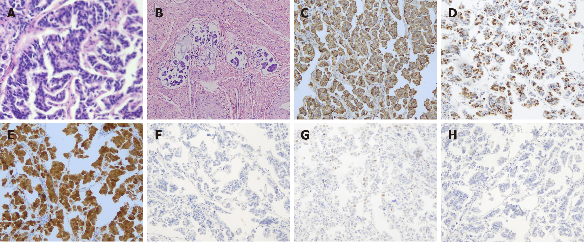
Figure 2 Cell clusters.
A: Pathological images of hematoxylin and eosin-stained samples showing high-grade serous carcinoma (H&E × 40); B: The micropapillary components form a large number of cancer emboli within the blood vessels; C: EMA; D: Ki67; E: P16 revealed diffusely positive staining (EnVision × 100); F: NapsinA; G: P5; H: WT1 all yielded negative results (EnVision × 100).
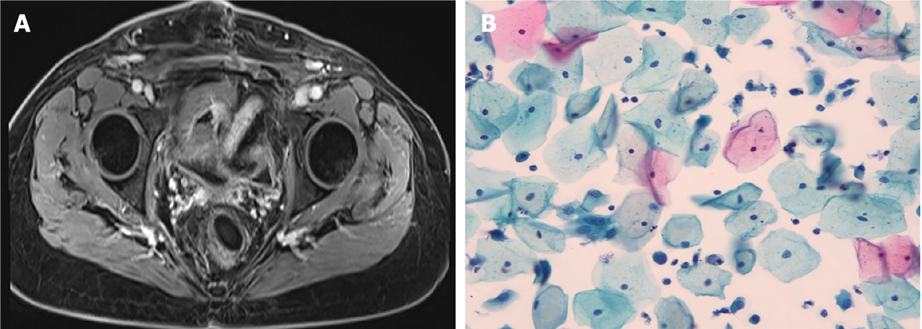
Figure 3 Images of pelvic magnetic resonance imaging and liquid-based cytology.
A: Images of pelvic magnetic resonance imaging with IV contrast scan when followed-up 53 months; B: Images of liquid-based cytology testing of the vaginal during the followed-up 53 months.
- Citation: Huang HQ, Yang L, Li QL, Sun CT, Gong FM. Human papillomavirus associated serous carcinoma of the uterine cervix in a patient with long-term survival: A case report. World J Clin Cases 2025; 13(22): 104643
- URL: https://www.wjgnet.com/2307-8960/full/v13/i22/104643.htm
- DOI: https://dx.doi.org/10.12998/wjcc.v13.i22.104643


