Copyright
©The Author(s) 2025.
World J Clin Cases. Aug 6, 2025; 13(22): 104165
Published online Aug 6, 2025. doi: 10.12998/wjcc.v13.i22.104165
Published online Aug 6, 2025. doi: 10.12998/wjcc.v13.i22.104165
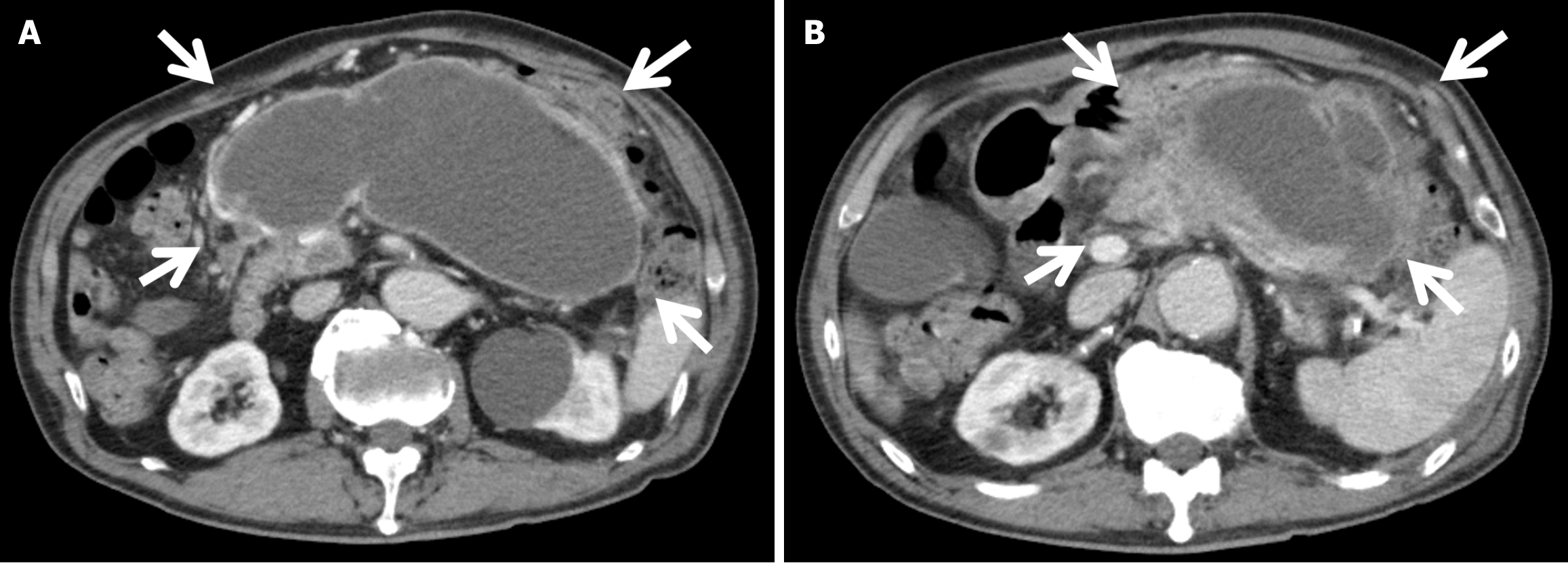
Figure 1 Contrast-enhanced computed tomography on postoperative day 42.
The heterogeneous enhancement and fluid collection (surrounded by white arrows) in the pancreatic body and tail, which were consistent with walled-off necrosis (WON). A: Caudal slice of the WON; B: Cranial slice of the WON.
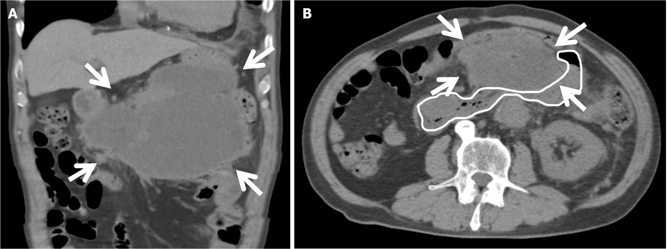
Figure 2 Computed tomography on postoperative day 42.
These computed tomography showed that the walled-off necrosis (arrows) was compressing the horizontal part of the duodenum (outlined in the white line). A: Coronal; B: Axial.
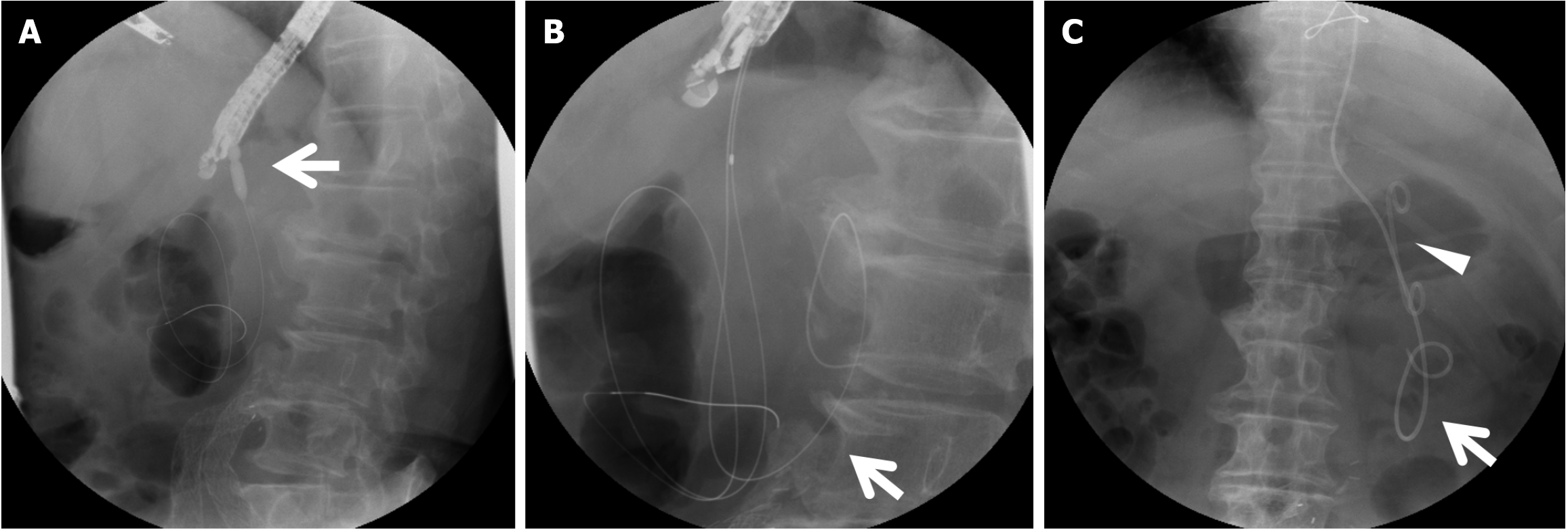
Figure 3 Endoscopic drainage of walled-off necrosis on postoperative day 50.
A: Dilation of the puncture site of a gastric wall using a dilation balloon (arrow); B: Additional wire (arrow) insertion; C: Double pig tail plastic stent (arrowhead) and pigtail nasal drainage tube (arrow) placement.
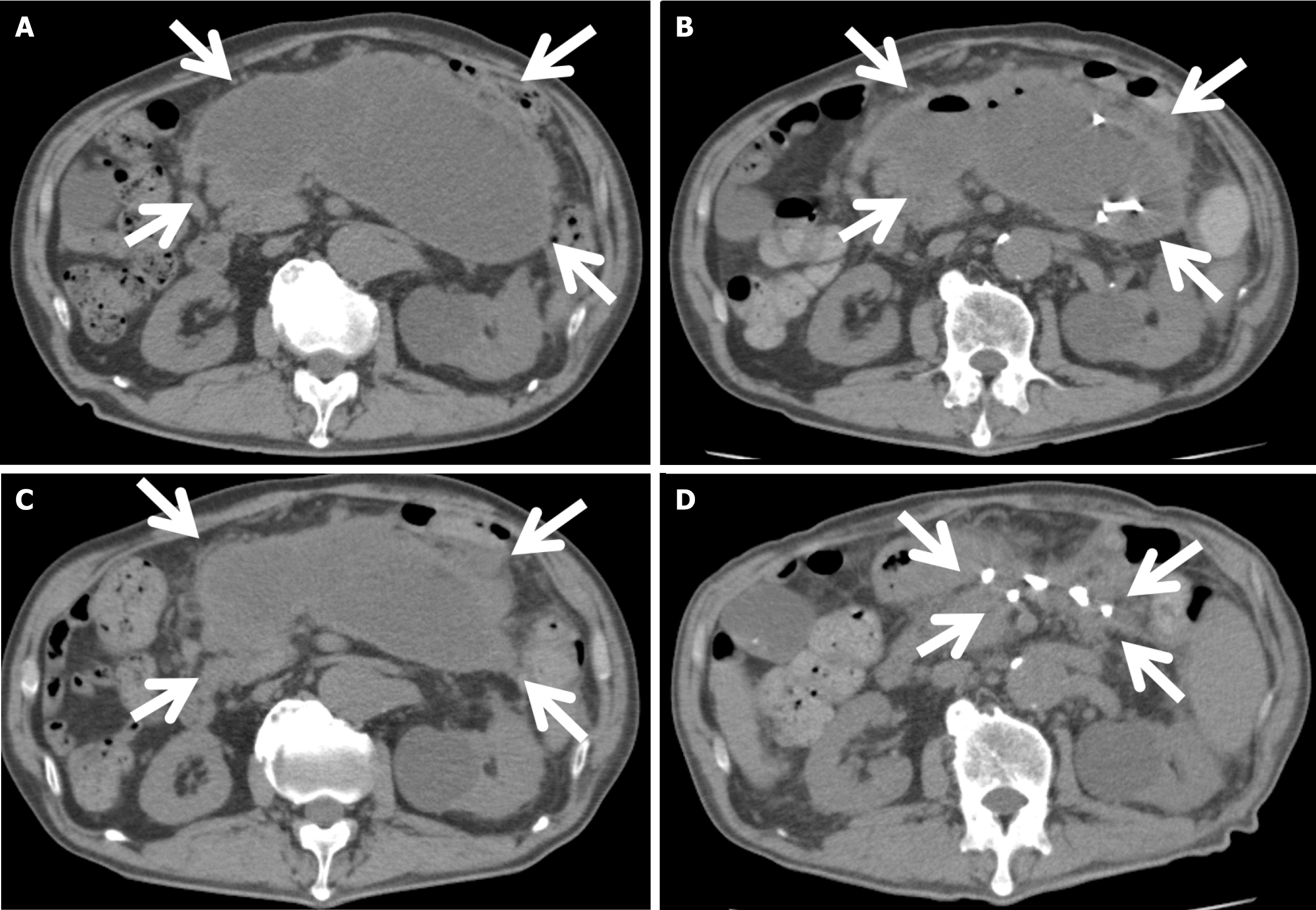
Figure 4 Abdominal computed tomography showing gradual shrinkage of walled-off necrosis.
A: On postoperative day 42 (before the drainage) the diameter of walled-off necrosis (WON) (surrounded by arrows) was 18.5 cm; B: On postoperative day 53 (3 days after the first drainage) the diameter of WON (surrounded by arrows) was 16 cm; C: On postoperative day 63 (13 days after the first drainage, before the second drainage) the diameter of WON (surrounded by arrows) was 18 cm; D: On postoperative day 165 (115 days after the first drainage, 78 days after the second drainage) WON (surrounded by arrows) almost disappeared.
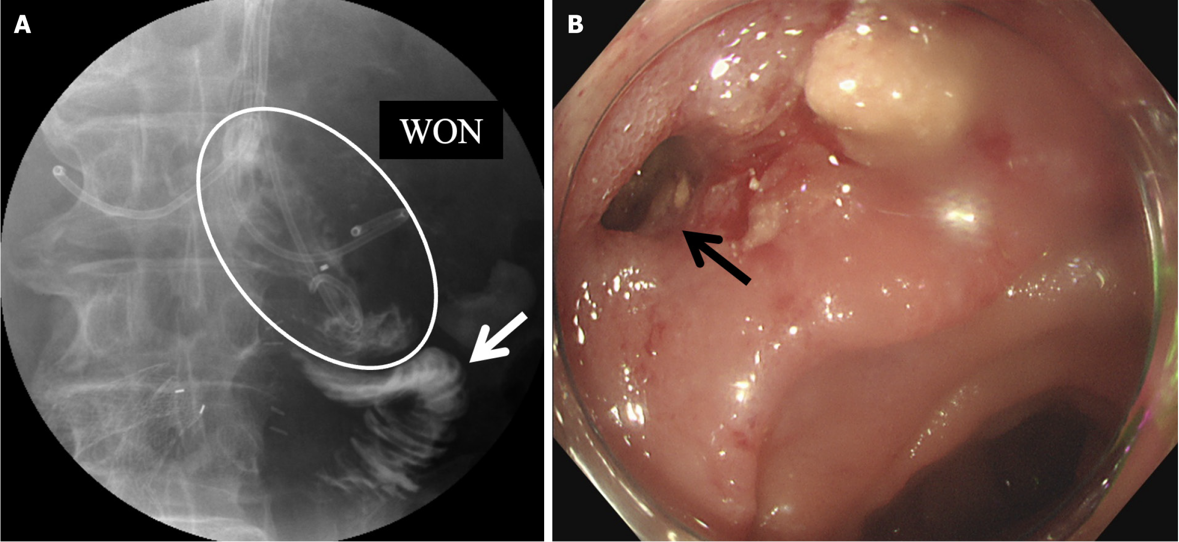
Figure 5 Image examination proved the fistula between walled-off necrosis and the duodenum.
A: The upper gastrointestinal tract contrast study showing a fistula between the walled-off necrosis (white circle) and duodenum (white arrow); B: Endoscopy showing the position of the fistula (black arrow). WON: Walled-off necrosis.
- Citation: Inoue Y, Yata Y, Yokota Y, Li ZL, Kawabata K. Acute pancreatitis after total aortic arch replacement leading to walled-off necrosis: A case report and review of literature. World J Clin Cases 2025; 13(22): 104165
- URL: https://www.wjgnet.com/2307-8960/full/v13/i22/104165.htm
- DOI: https://dx.doi.org/10.12998/wjcc.v13.i22.104165


