Copyright
©The Author(s) 2025.
World J Clin Cases. Jul 16, 2025; 13(20): 105133
Published online Jul 16, 2025. doi: 10.12998/wjcc.v13.i20.105133
Published online Jul 16, 2025. doi: 10.12998/wjcc.v13.i20.105133
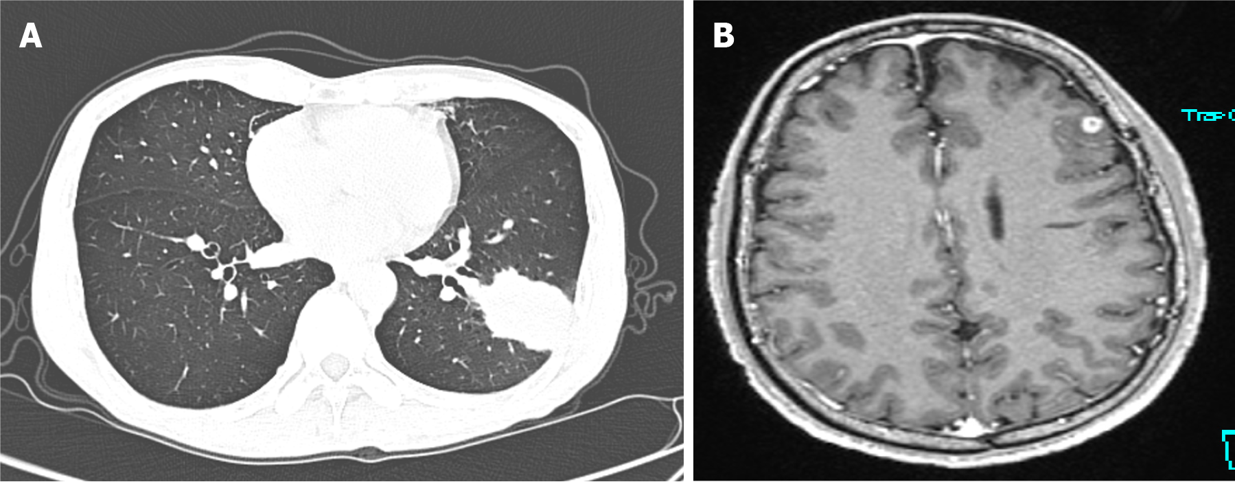
Figure 1 Imaging examinations.
A: Chest computed tomography (December 31, 2020) showing a wedge-shaped high-density lesion in the lower lobe of the left lung; B: Gd-enhanced magnetic resonance imaging (December 2020) showing a small round enhancing lesion in the left frontal lobe.
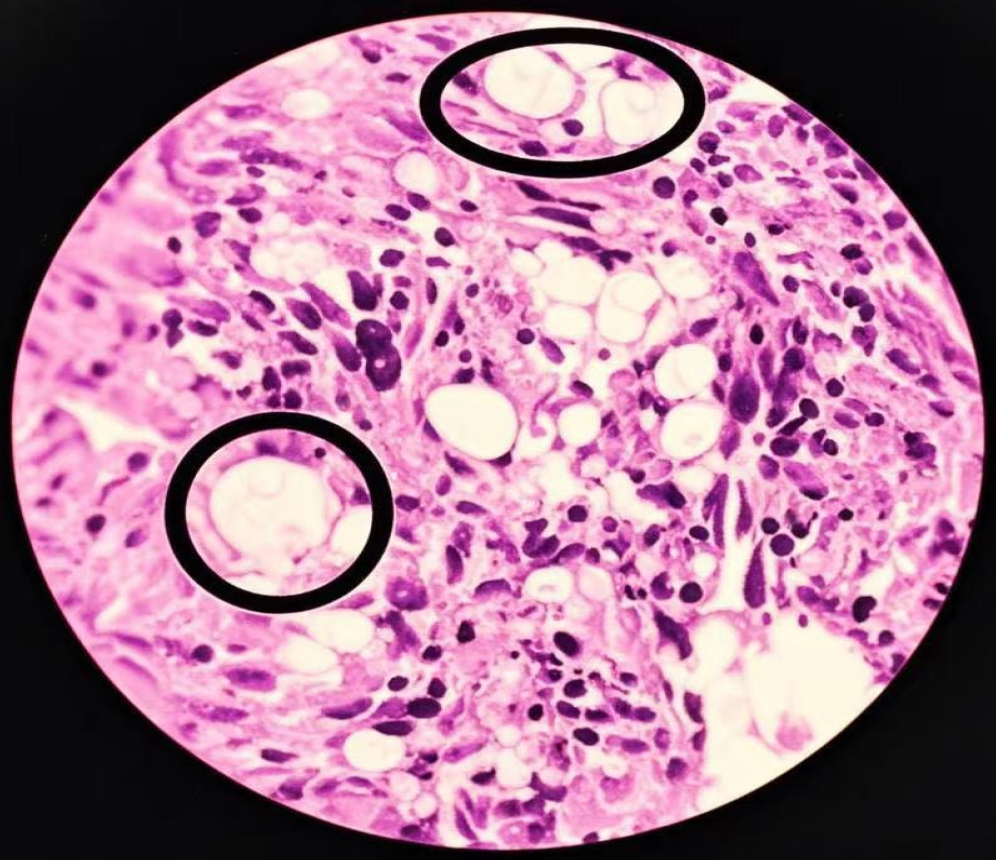
Figure 2 Histopathological features of the left lung mass (hematoxylin and eosin stain; × 400).
Large “titan cell” pathogens are evident (circles) with budding of smaller pathogens surrounded by transparent halos. These are accompanied by monocyte-macrophage infiltration with fibrosis, necrosis and granuloma formation. Under low magnification, the lung tissue shows chronic inflammatory changes, and granulomatous nodules formed by the aggregation of histiocytes. Epithelioid cells and multinucleated giant cells are observed, accompanied by fibrous tissue hyperplasia and lymphocyte infiltration. Under high magnification, round and translucent bodies can be seen in the alveolar cavity and multinucleated giant cells. The centers of these bodies are lightly stained, with thin walls, and a clear halo can be seen around them. Combined with the results of special staining, it is consistent with cryptococcus infection.
Figure 3 Follow-up computed tomography chest and brain magnetic resonance imaging after secondary reintroduction of flucytosine and amphotericin B.
The lung lesion showed considerable improvement however new brain lesions had formed with some increase in size of the initial cerebral mass.
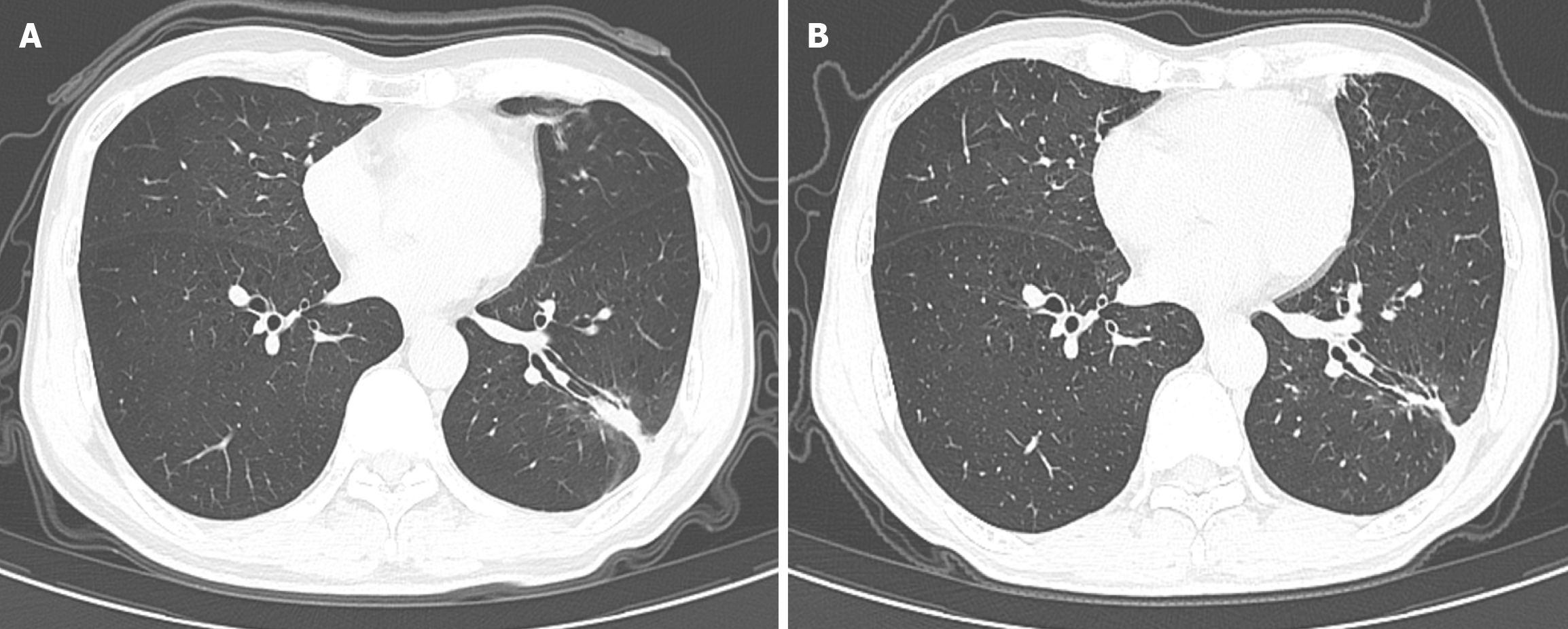
Figure 4 Follow-up computed tomography scans of the thorax showing near resolution of the lung mass (arrows).
A: Image was reported 12 months after initiation of antifungal therapy; B: Image a further 22 months later.
- Citation: Gao CY, Yang XJ, Guo E, Zheng YL. Invasive pulmonary cryptococcosis mimicking metastatic lung cancer: A case report and review of literature. World J Clin Cases 2025; 13(20): 105133
- URL: https://www.wjgnet.com/2307-8960/full/v13/i20/105133.htm
- DOI: https://dx.doi.org/10.12998/wjcc.v13.i20.105133


