Copyright
©The Author(s) 2025.
World J Clin Cases. Jul 16, 2025; 13(20): 100169
Published online Jul 16, 2025. doi: 10.12998/wjcc.v13.i20.100169
Published online Jul 16, 2025. doi: 10.12998/wjcc.v13.i20.100169
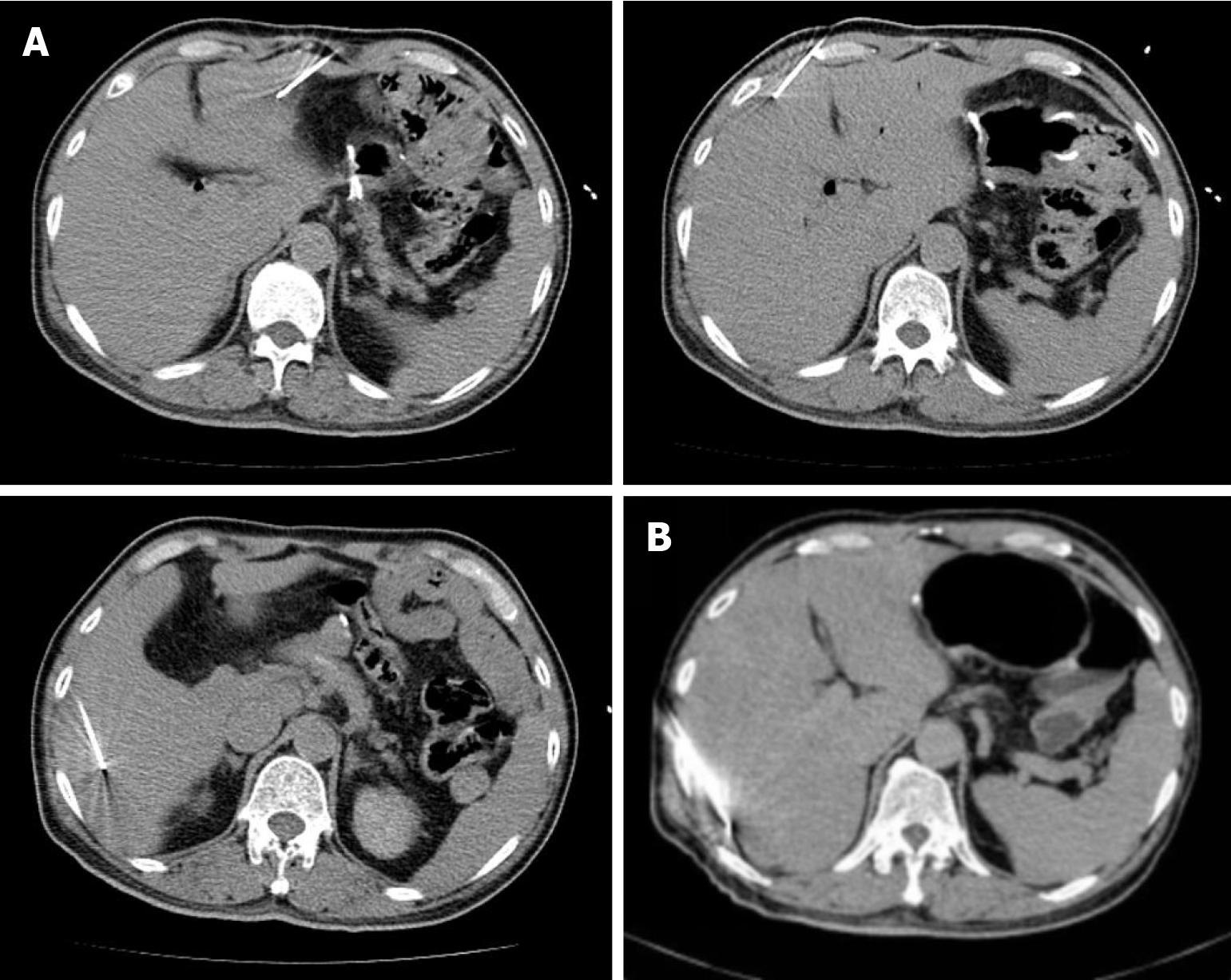
Figure 1 Radiofrequency ablation of liver metastases was performed on the patient.
A: Radiofrequency ablation of the left outer lobe, the right anterior lobe, and the right upper lobe of the liver was performed under computed tomography guidance, and the procedure went smoothly; B: On March 20, 2020, the patient underwent radiofrequency ablation of liver metastases.
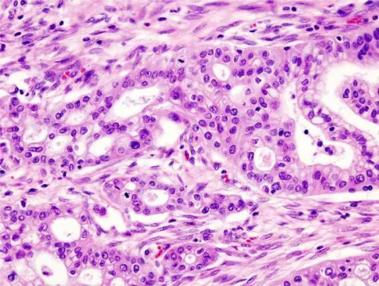
Figure 2
The postoperative pathological report of the patient.
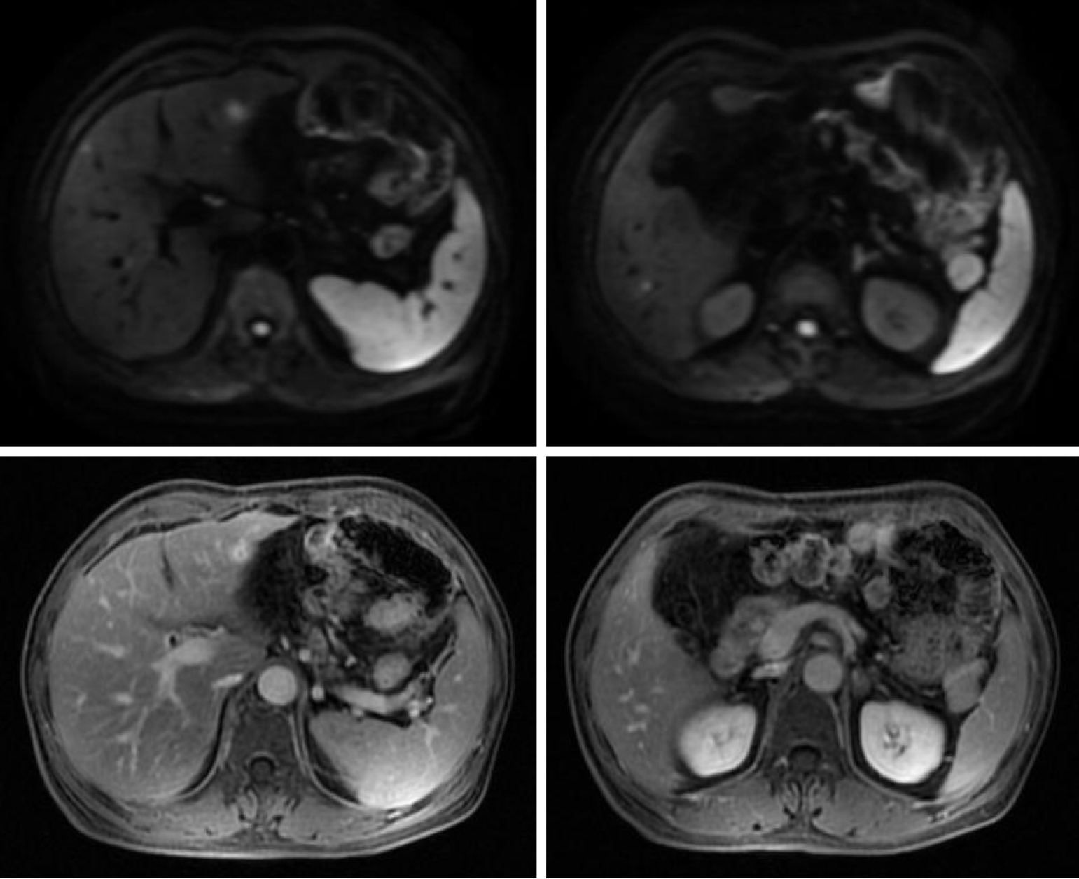
Figure 3 Original pancreatic cancer after surgery and chemotherapy.
Postoperative changes in the head of the pancreas: no clear abnormal enhancement foci are observed in the operation area. Follow-up combined with the examination of tumor markers is recommended. Abnormal enhancement signals in the left outer lobe of the liver and metastatic foci are visible. Speckled and abnormal enhancement foci in the upper part of the right anterior lobe of the liver, a possible hemangioma. Metastatic tumors were not excluded for the time being. Small cyst in the right lobe of the liver. Gallbladder agenesis. Parasplenic incidental observation: small adenoma of the left adrenal gland. Small adenoma of the left adrenal gland. Small adenoma of the left adrenal gland.
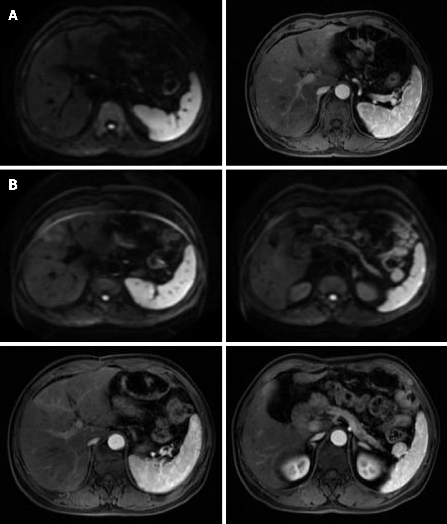
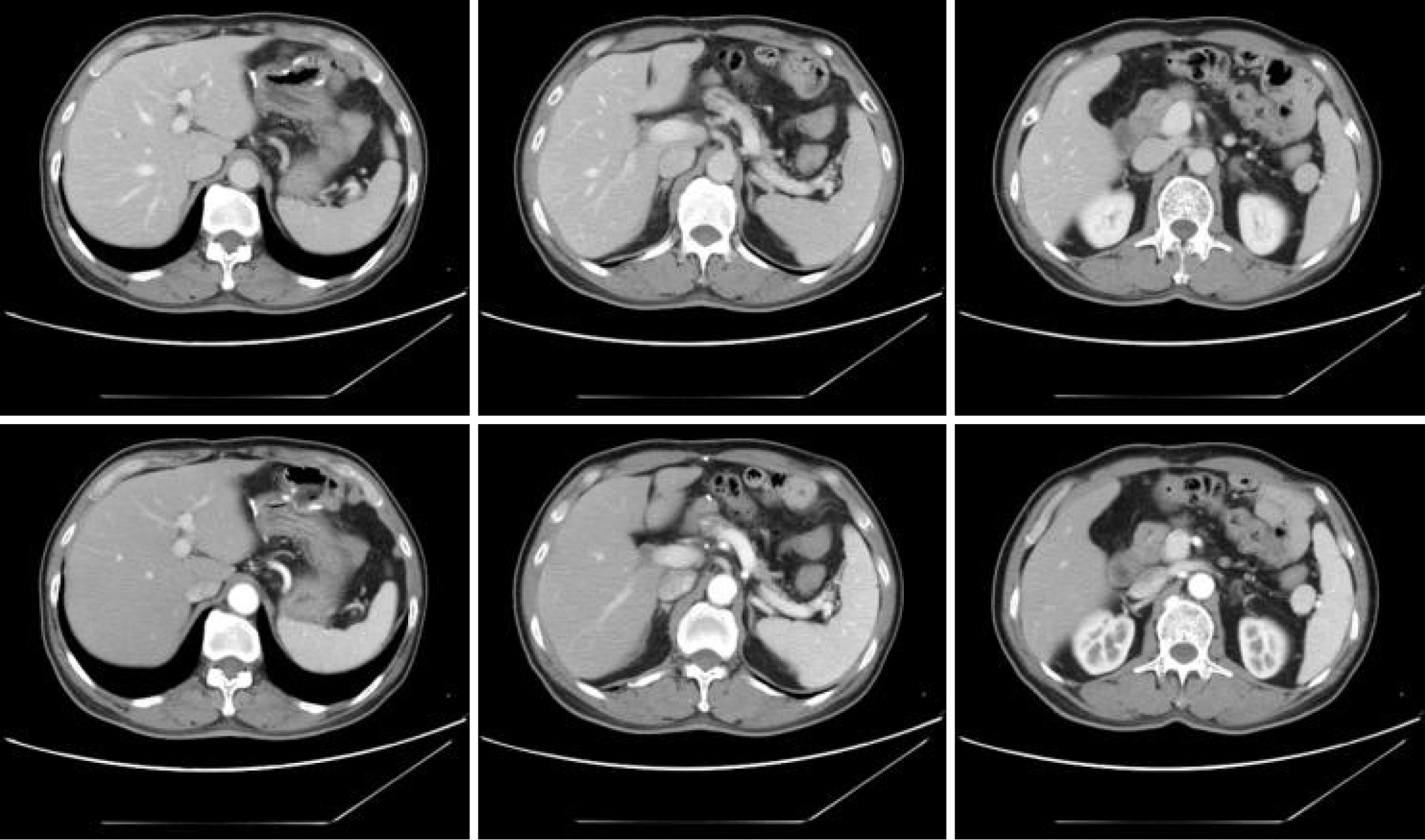
Figure 4 Original pancreatic cancer liver metastases after radiofrequency ablation.
A: Postoperative changes in the head of the pancreas: no clear abnormal enhancement foci are observed in the operated area, but the central pancreatic duct is mildly dilated. It is recommended to follow up for review in combination with tumor markers. Minor nodular enhancement in the arterial stage of the left inner and outer lobes of the liver and iso-signals in the hepatic and biliary stages; pseudoenhancement was considered. No enhancement signals in the left inner lobe of the liver or in the right posterior lobe of the liver, which was false enhancement. Absence of the gallbladder; Parasplenic incidental observations: Small left adrenal adenoids. Postradiofrequency changes, compared with those in the July 07, 2020 slices: Part of the lesion was slightly reduced. The gallbladder is missing. Parasplenic incidental observations: small adenoma of the left adrenal gland; B: Pancreatic head postoperative changes: no clear abnormal enhancement foci in the operated area. Abnormal enhancement foci in the upper part of the right posterior lobe of the liver, which is a newly-emerged lesion, and it is recommended to follow up and review the disease. The left inner and outer lobes of the liver and the right posterior lobe have no enhancement signals, which is a postoperative change of the radiofrequency operation; compared with the January 12, 2019.13 slices, the lesion has shrunk. The left inner lobe lesion has no significant change in comparison with the previous slices. The gallbladder is absent. Parasplenic incidental observations: small adenoma of the left adrenal gland.
Figure 5
Return to the hospital for review in March 2024 showing stabilization.
- Citation: Yong JP, Mu XY, Zhou CF, Zhang KK, Gao JQ, Guo ZZ, Zhou SF, Ma Z. Radiofrequency ablation of liver metastases in a patient with pancreatic cancer and long-term survival: A case report. World J Clin Cases 2025; 13(20): 100169
- URL: https://www.wjgnet.com/2307-8960/full/v13/i20/100169.htm
- DOI: https://dx.doi.org/10.12998/wjcc.v13.i20.100169


