Copyright
©The Author(s) 2025.
World J Clin Cases. Jan 16, 2025; 13(2): 97834
Published online Jan 16, 2025. doi: 10.12998/wjcc.v13.i2.97834
Published online Jan 16, 2025. doi: 10.12998/wjcc.v13.i2.97834
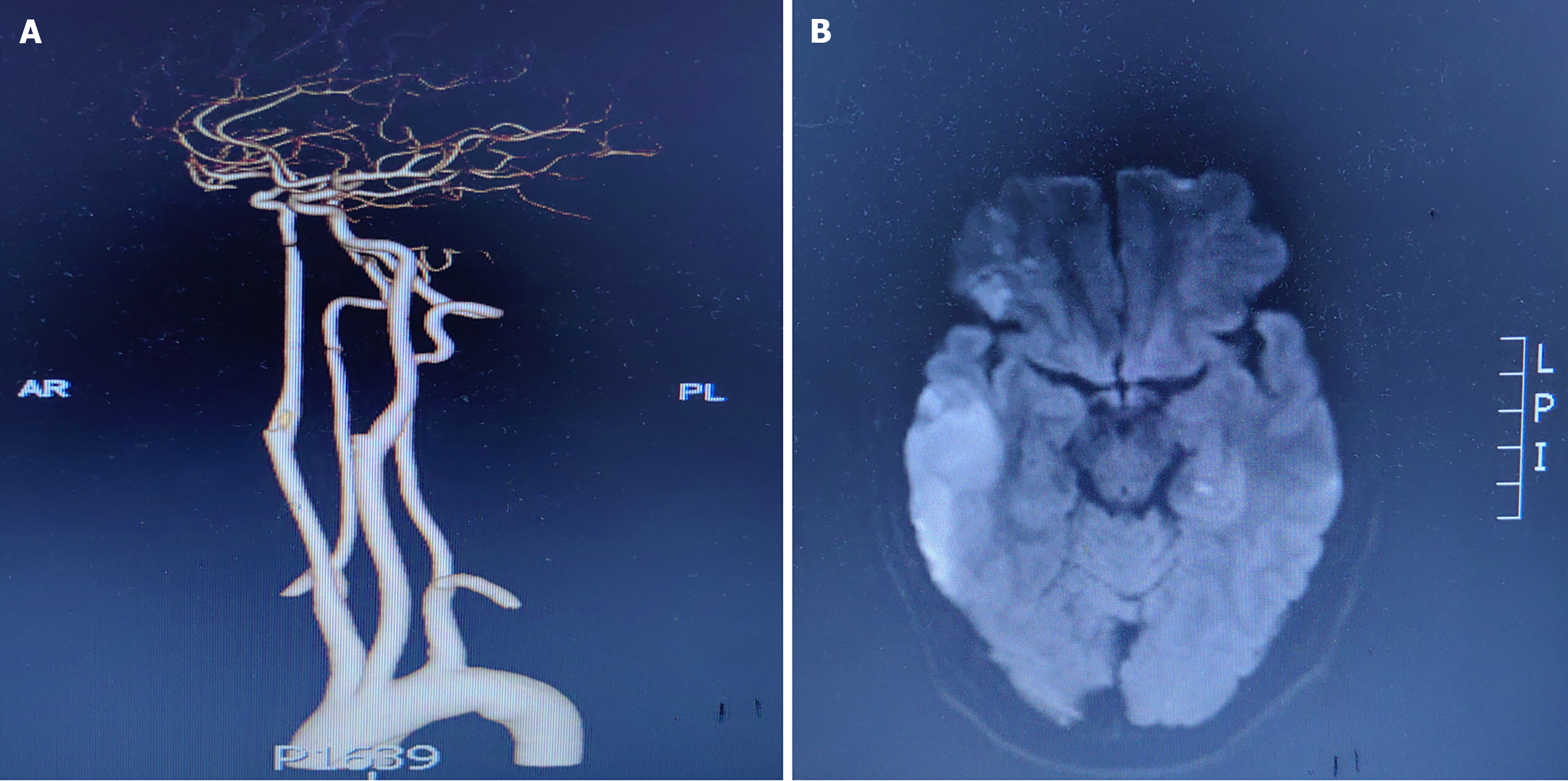
Figure 1 Imaging examinations.
A: Computed tomography angiography showed fat embolism in the proximal right internal carotid artery and proximal right external carotid artery; B: Diffusion-weighted imaging showed high signal intensity in the left cerebral hemisphere.
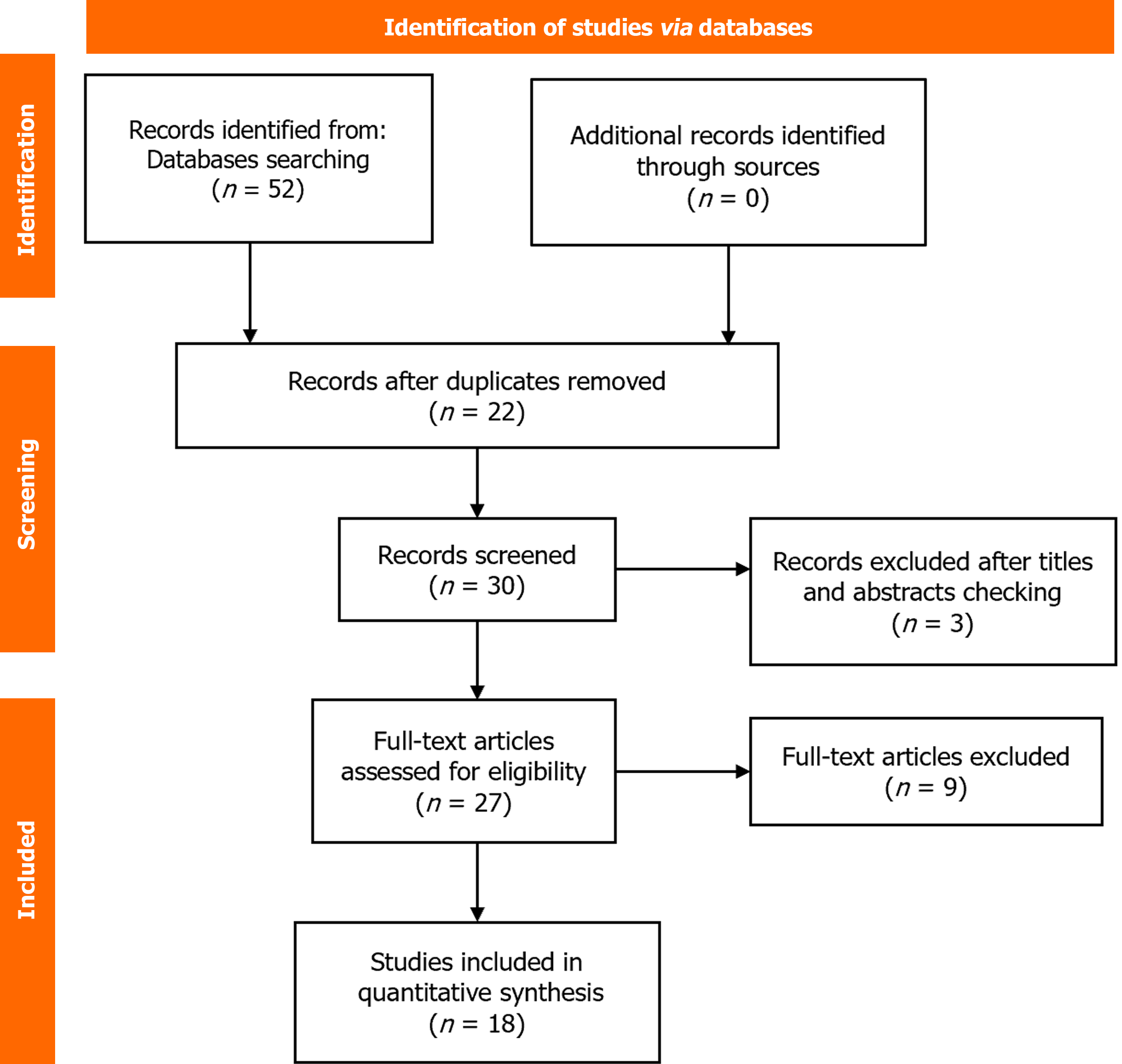
Figure 2 Literature search process.
- Citation: Chen XY, Shen F, Cheng C, Wang YH, Cheng WC, Yuan DZ, Huang W. Cerebral fat embolism following autologous fat injection in facial reconstruction: A case report. World J Clin Cases 2025; 13(2): 97834
- URL: https://www.wjgnet.com/2307-8960/full/v13/i2/97834.htm
- DOI: https://dx.doi.org/10.12998/wjcc.v13.i2.97834


