Copyright
©The Author(s) 2025.
World J Clin Cases. Jul 6, 2025; 13(19): 103844
Published online Jul 6, 2025. doi: 10.12998/wjcc.v13.i19.103844
Published online Jul 6, 2025. doi: 10.12998/wjcc.v13.i19.103844
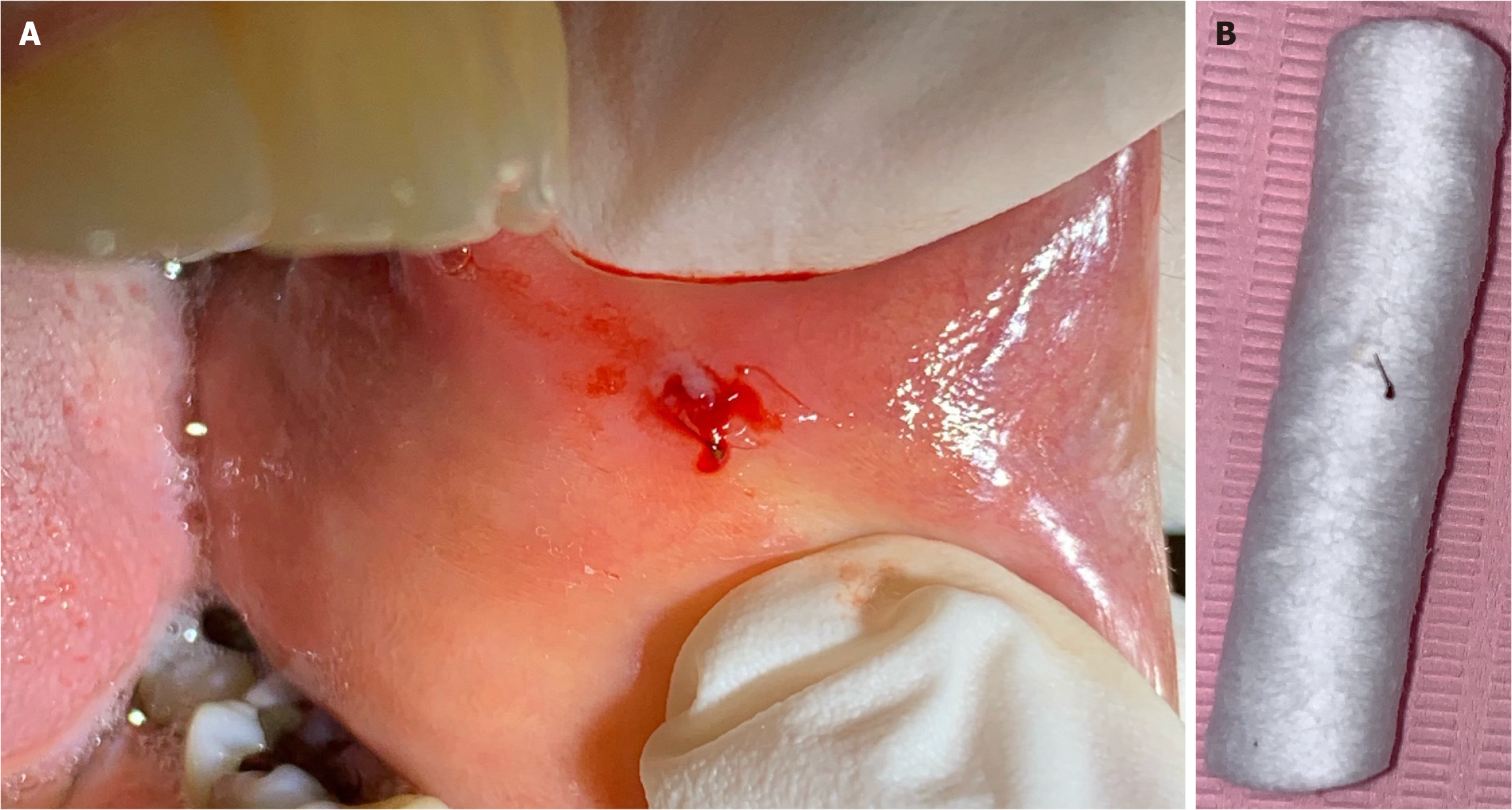
Figure 1 Clinical photos of the honeybee stinger.
A: A clinical intraoral photo of a 2-mm black, long, thin foreign body came out with the pus and blood from the vesicle on the left buccal mucosa, B: A honeybee stinger on a cotton roll.
Figure 2 A microscopic photo of the foreign body confirmed a honeybee stinger.
- Citation: Aloyouny AY, Albagieh HN, Aleyoni R, Jammali G, Alhuzali K. Unusual foreign body in the buccal mucosa: A case report. World J Clin Cases 2025; 13(19): 103844
- URL: https://www.wjgnet.com/2307-8960/full/v13/i19/103844.htm
- DOI: https://dx.doi.org/10.12998/wjcc.v13.i19.103844


