Copyright
©The Author(s) 2025.
World J Clin Cases. Jul 6, 2025; 13(19): 103585
Published online Jul 6, 2025. doi: 10.12998/wjcc.v13.i19.103585
Published online Jul 6, 2025. doi: 10.12998/wjcc.v13.i19.103585
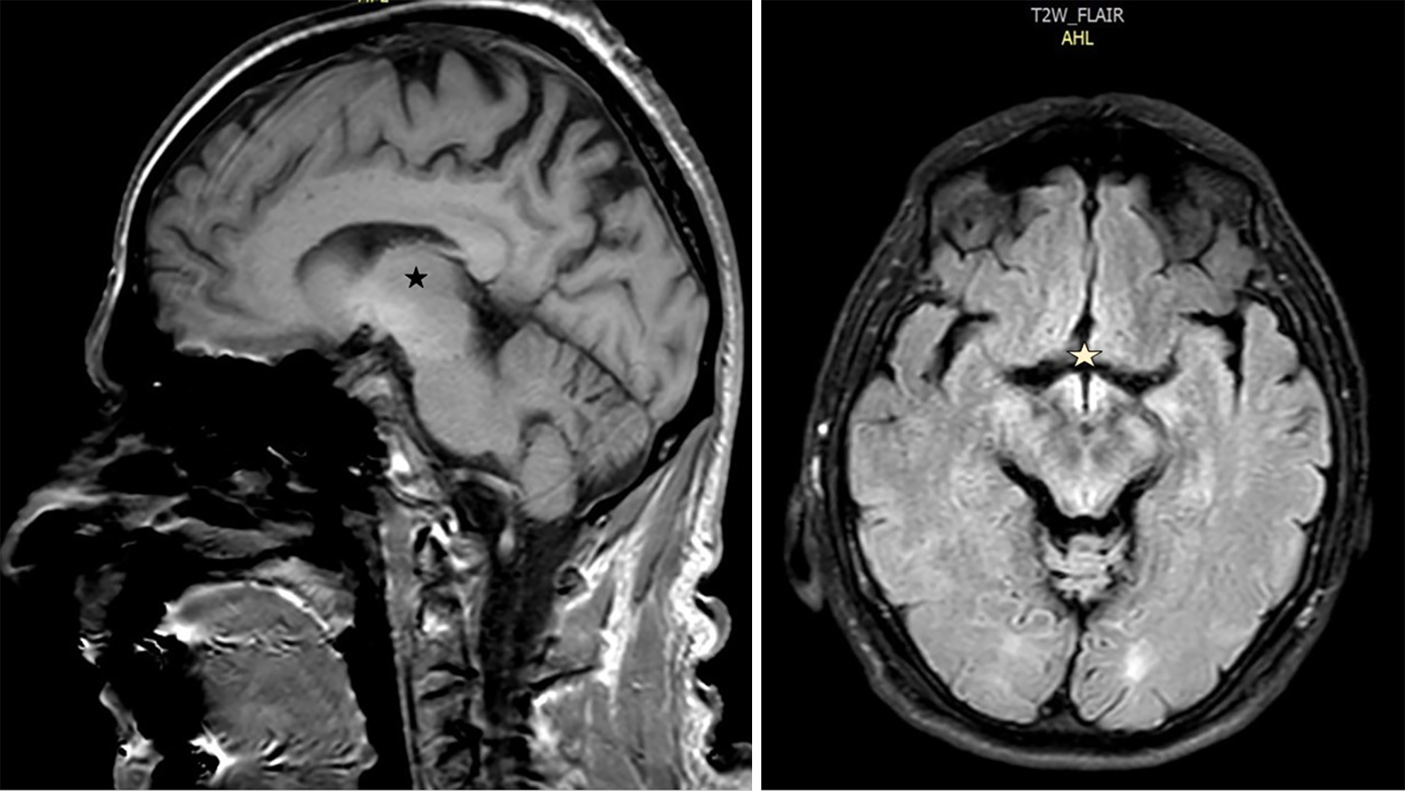
Figure 1 Magnetic resonance imaging of the head showing hyperintense signals in the fluid attenuation inversion recovery sequence, involving hypothalamic structures (mammillary bodies, right inset), subthalamic and adjacent midbrain areas.
Left inset: Sagittal images (black star); right inset: Axial images (yellow star).
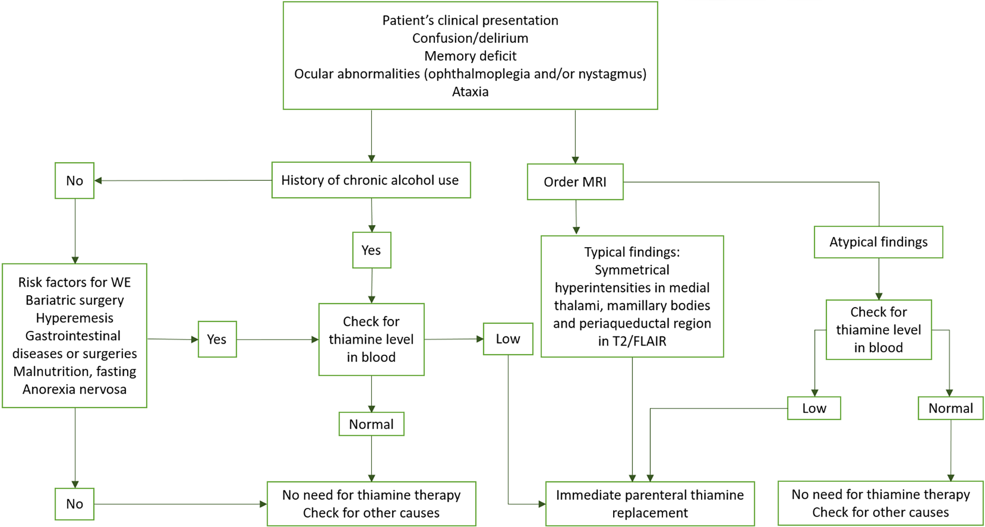
Figure 2 Algorithm: Practical diagnostic and therapeutic approach to a patient with Wernicke encephalopathy.
FLAIR: Fluid attenuation inversion recovery; MRI: Magnetic resonance imaging; WE: Wernicke encephalopathy.
- Citation: Roçi E, Mara E, Dodaj S, Vyshka G. Wernicke encephalopathy presenting as a stroke mimic: A case report. World J Clin Cases 2025; 13(19): 103585
- URL: https://www.wjgnet.com/2307-8960/full/v13/i19/103585.htm
- DOI: https://dx.doi.org/10.12998/wjcc.v13.i19.103585


