Copyright
©The Author(s) 2025.
World J Clin Cases. Jul 6, 2025; 13(19): 102212
Published online Jul 6, 2025. doi: 10.12998/wjcc.v13.i19.102212
Published online Jul 6, 2025. doi: 10.12998/wjcc.v13.i19.102212
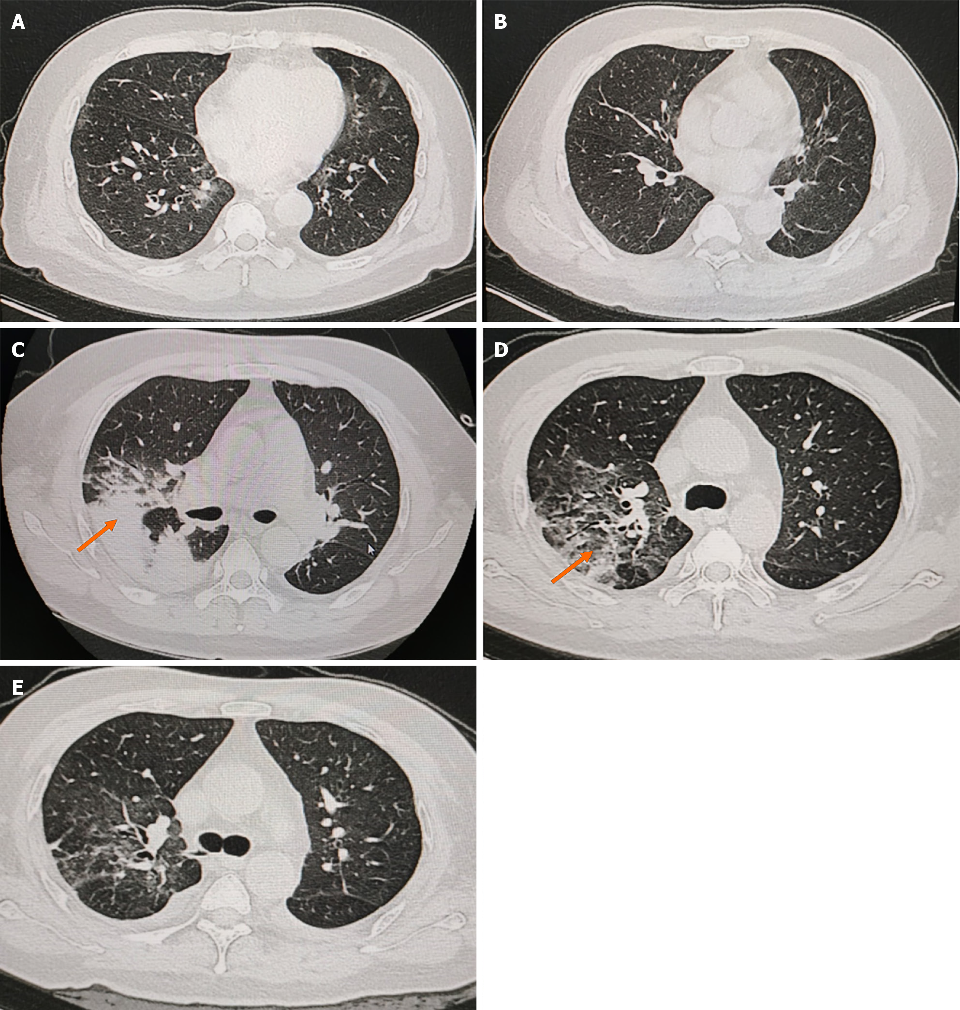
Figure 1 Computed tomography imaging images at the time of initial hospitalization and disease progression.
A and B: High-resolution computed tomography (CT) images to consider viral infections in both lungs; C: Pulmonary artery imaging on March 23, 2023, reveals multiple inflammations in both lungs are significantly more advanced than before. CT angiography imaging of the pulmonary arteries reveals no significant abnormalities. The orange arrow shows prominent solid exudative changes in the right upper lung; D: Multiple inflammations in both lungs are significantly more resorbed than before; E: High-resolution CT of the lungs, performed on April 13, 2023, demonstrates partial resolution of the lesions in the upper and dorsal segments of the right upper and lower lobes, along with a small amount of bilateral pleural fluid effusion. Moreover, partial distension of the adjacent lower lungs is observed.
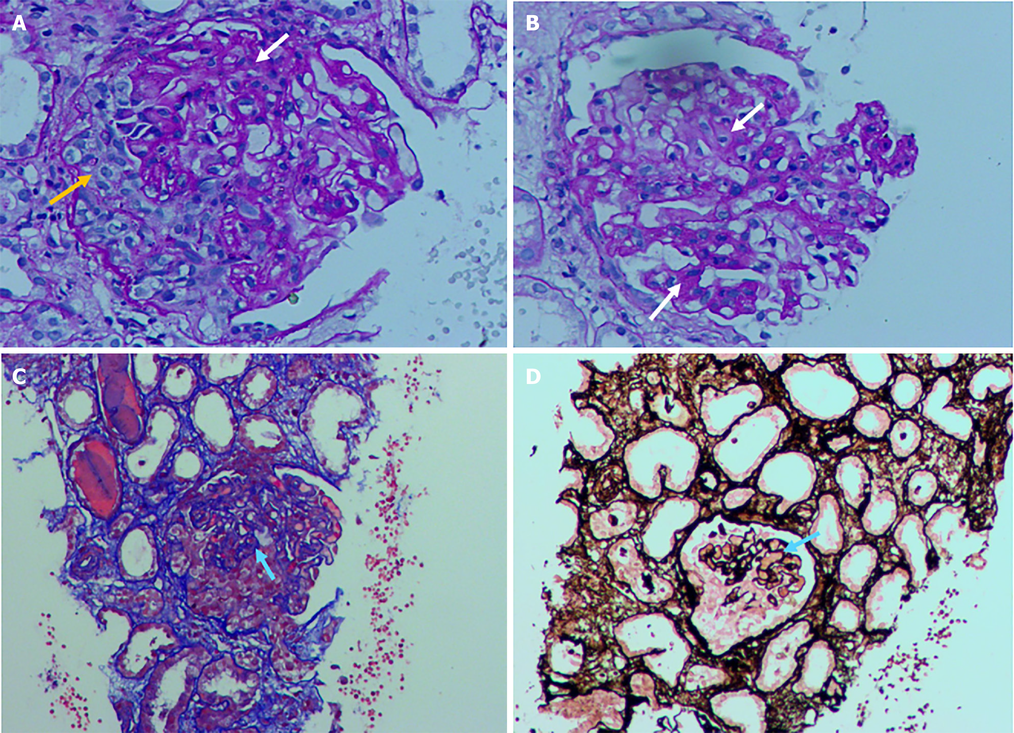
Figure 2 Light microscopic picture of renal pathology.
The light microscopy specimen in this case reveals the presence of five glomeruli, including three marginal glomeruli with crescentic lesions (3/5), while only two intact glomeruli are observed. PASM-Masson staining: Glomeruli with suspected erythrophilic deposits, segments with suspicious “pegs”. Renal tubular interstitium with severe chronic lesions, multiple small foci of tubular atrophy, basement membrane thickening, some tubular epithelial cells with large nuclei, and some cells with intranuclear inclusion bodies. Focal renal tubular epithelial cells with brush border detachment, some renal tubular epithelial cells with fine apparent granular degeneration and vacuolar degeneration, and tubular lumen with visible protein tubular pattern. Interstitial fibrosis and edema, multifocal infiltration of single nucleated cells, and eosinophils are seen in small foci. Segmental or total hyaline degeneration of small arteries. A and B: Periodic acid-Schiff stain of glomeruli, fibrous necrosis and crescent formation can be seen; C: Masson-Trichrome, glomeruli with suspected erythrophilic deposits, segments with suspicious “pegs”; D: Periodic-acid silver methenamine. White arrows: Fibrinoid necrosis; yellow arrows: Fibrocellular crescent; blue arrow: Basement membrane thickening.
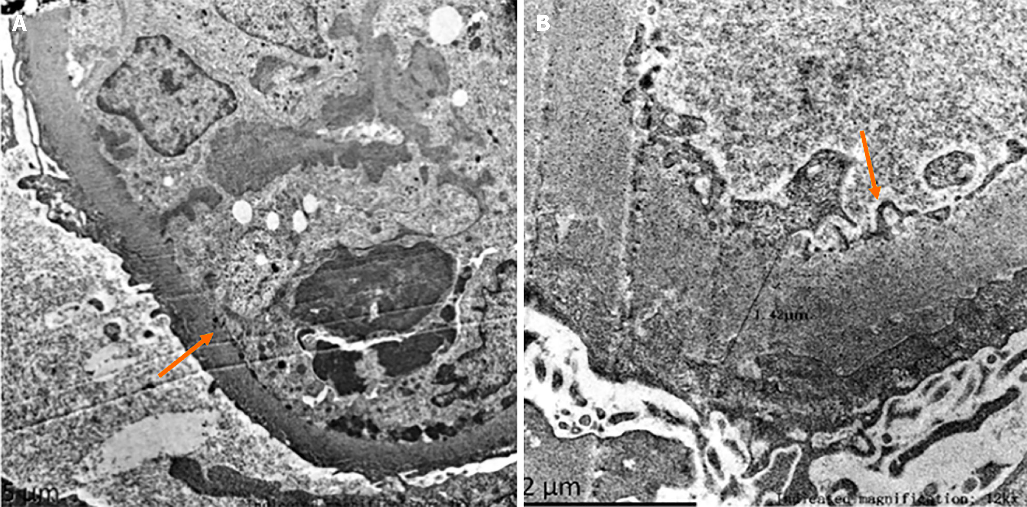
Figure 3 Electron microscopic picture of renal pathology.
Electron microscopy reveals mild to moderate glomerular mesangial hyperplasia accompanied by segmental endothelial cell proliferation and neutrophil infiltration. Multiple immune complexes are identified, along with diffuse basement membrane thickening, the formation of small pegs, extensive fusion of pedicles, and the characteristic double-tracking and layering pattern. Multifocal renal tubular atrophy is observed, along with edema, vacuolar degeneration in some tubular epithelial cells, interstitial edema, focal fibrous tissue hyperplasia, and scattered inflammatory cell infiltration. A: Diffuse basement membrane thickening (orange arrow); B: The formation of small pegs (orange arrow).
- Citation: Li MR, Li LY, Tang J, Sun J. Chronic hepatitis B triggering antineutrophil cytoplasmic antibody-associated vasculitis complicated by glomerulonephritis: A case report. World J Clin Cases 2025; 13(19): 102212
- URL: https://www.wjgnet.com/2307-8960/full/v13/i19/102212.htm
- DOI: https://dx.doi.org/10.12998/wjcc.v13.i19.102212


