Copyright
©The Author(s) 2025.
World J Clin Cases. Jun 26, 2025; 13(18): 103571
Published online Jun 26, 2025. doi: 10.12998/wjcc.v13.i18.103571
Published online Jun 26, 2025. doi: 10.12998/wjcc.v13.i18.103571
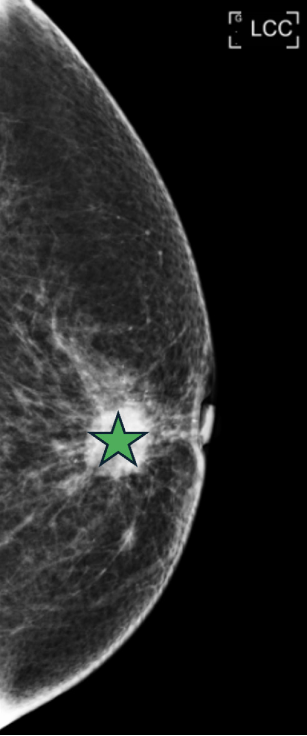
Figure 1 Mammography on February 6, 2023.
Well circumscribed lobular masses with pleomorphic calcifications in the left breast (green star, size: Up to about 1.78 cm, in the 12 o'clock, M/3, about 1.05 cm away from the nipple on the craniocaudal view view), probably neoplasms.
Figure 2 Breast sonography on February 6, 2023.
Multiple masses (yellow star at the left picture) with poorly defined margin and dilated duct in left breast, the biggest one (red star at the right picture) is at 12 o'clock position of left breast, (12/2 cm), 22.6 mm × 12.6 mm in size, might be neo-growth. Breast imaging reporting and data system category 4c: Moderate suspicious abnormality.
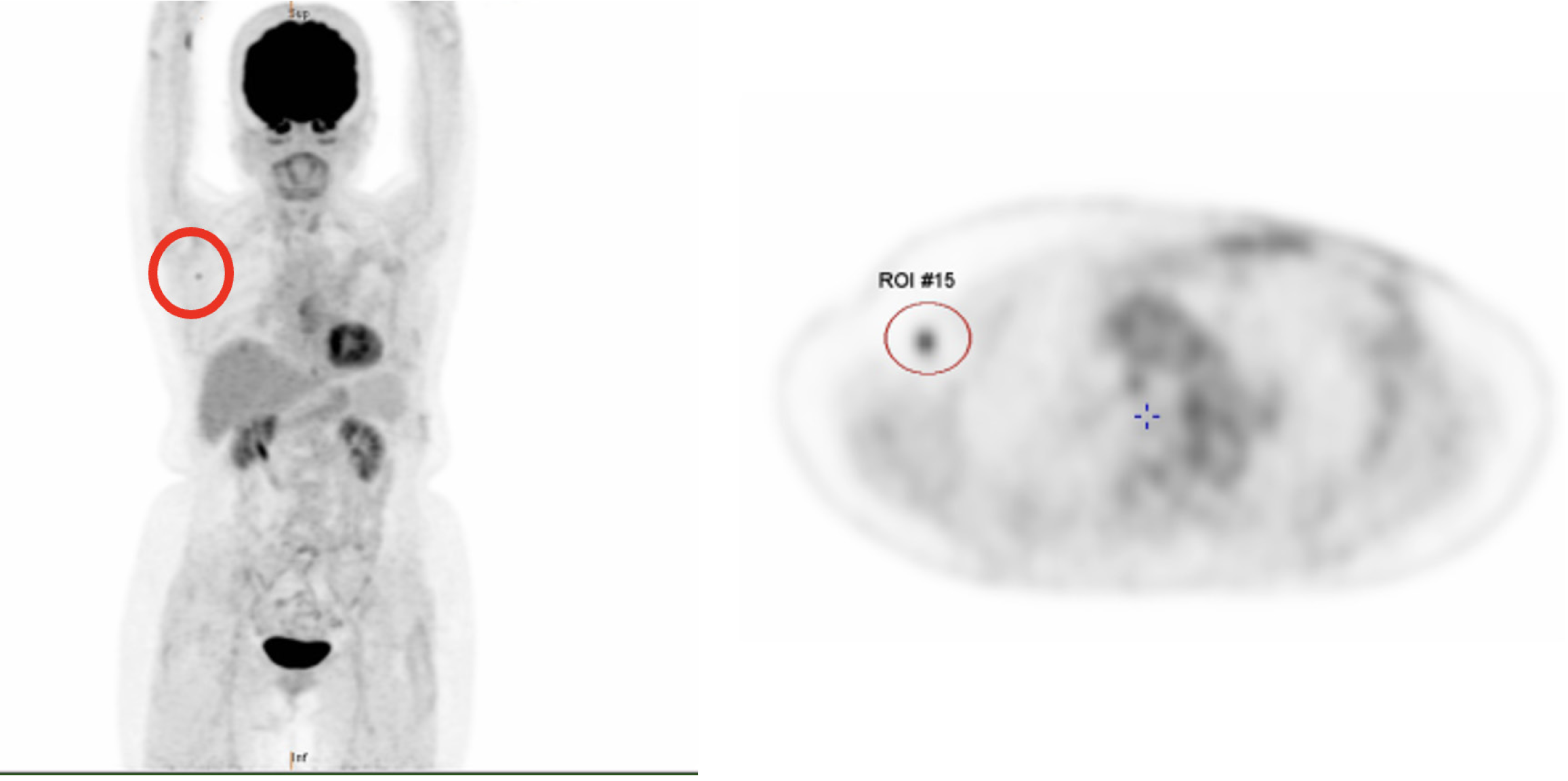
Figure 3 Fluorodeoxyglucose- positron emission tomography whole body scan on March 21, 2023 (coronal view and axial view).
One mild fluorodeoxyglucose-avidity over the right axillary region (red circle, maximal standard uptake value = 3.0, size > 0.4 cm) favoring reactive node. Based on clinical history [pT2(m)N0] and the imaging findings, the current PET finding suggesting no metabolic evidence of malignancy (left breast cancer), cT2(m)N0M0[35].
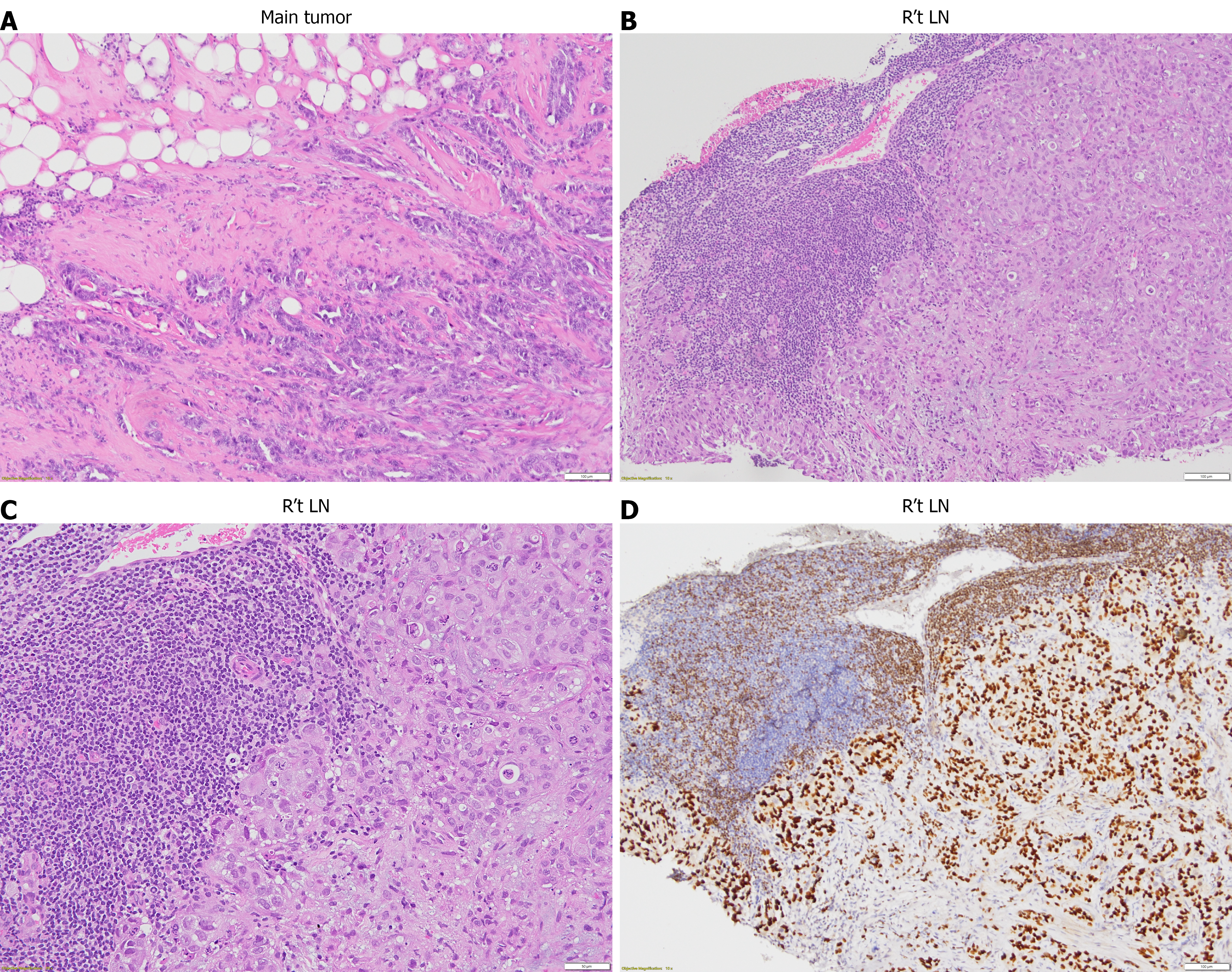
Figure 4 H&E stain of the main tumor and the right axillary lymph node and immunohistochemical stain of the right axillary lymph node.
A: H&E stain of the main tumor with magnification 100 ×. An invasive carcinoma consists of tumor cells with nuclear pleomorphism and high N/C ratio, arranged in distorted glandular to solid nested patterns, infiltrating to the fibrous stroma and adipose tissue; B and C: H&E stain of the right axillary lymph node with magnification 100 × (B) and 200 × (C). Follicular structure of normal lymphoid tissue is effaced by solid nested tumor cells with nuclear pleomorphism; D: Immunohistochemical stain of the right axillary lymph node with magnification 100 ×. Mammary origin of metastatic tumor is identified with positive GATA3. R’t LN: Right lymph node.
- Citation: Lin YT, Hong ZJ, Liao GS, Dai MS, Chao TK, Tsai WC, Sung YK, Chiu CH, Chang CK, Yu JC. Unexpected contralateral axillary lymph node metastasis without ipsilateral involvement in triple-negative breast cancer: A case report and review of literature. World J Clin Cases 2025; 13(18): 103571
- URL: https://www.wjgnet.com/2307-8960/full/v13/i18/103571.htm
- DOI: https://dx.doi.org/10.12998/wjcc.v13.i18.103571


