Copyright
©The Author(s) 2025.
World J Clin Cases. Jun 26, 2025; 13(18): 103438
Published online Jun 26, 2025. doi: 10.12998/wjcc.v13.i18.103438
Published online Jun 26, 2025. doi: 10.12998/wjcc.v13.i18.103438
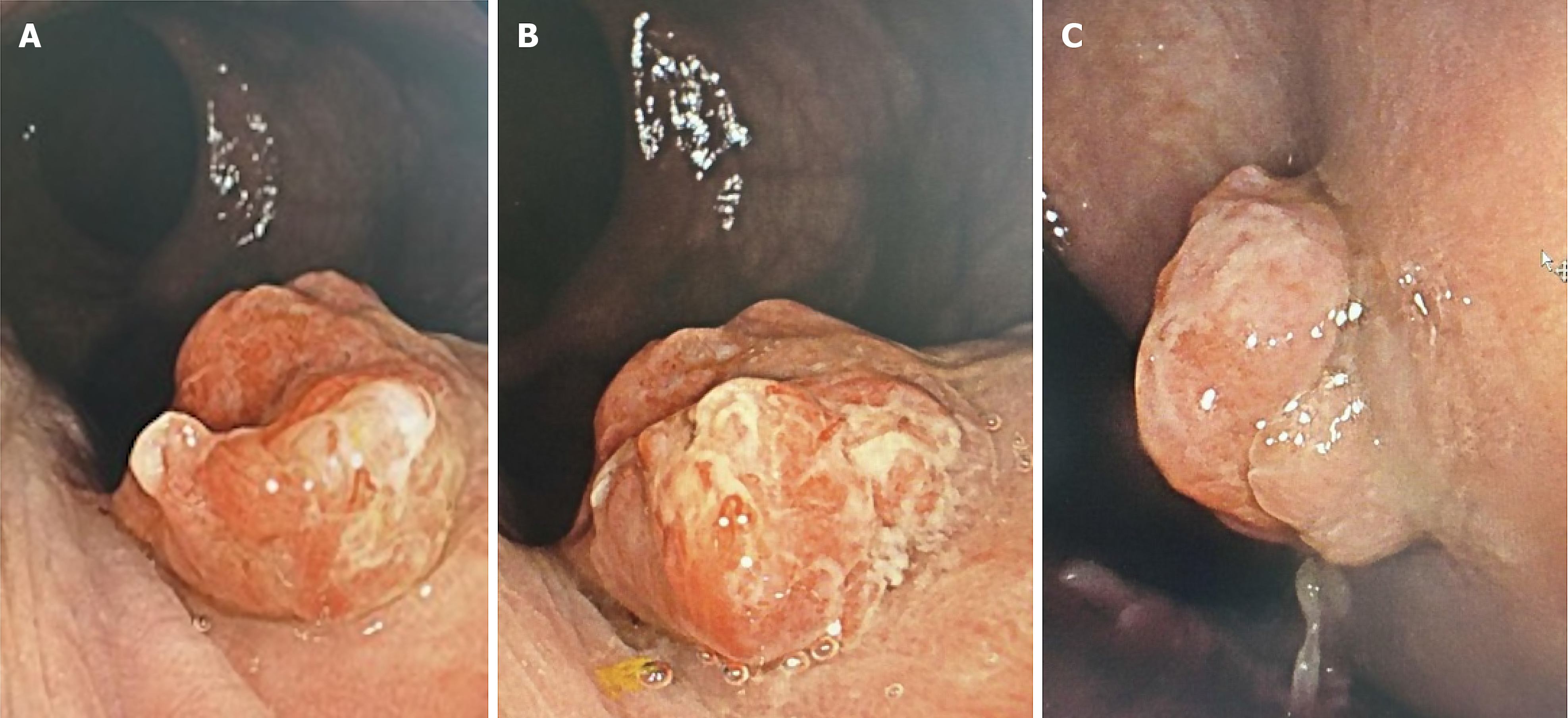
Figure 1 Erythematous, exophytic lesion in the distal rectum below the distal valve of Houston, proximal to the dentate line, measuring 13 mm × 10 mm, with an inflammatory, lobulated, and villous appearance, not involving more than 20% of the circumference or causing stenosis of the rectal lumen.
A: Frontal view of the polyp; B: Lateral view of the polyp; C: Medial view of the polyp.
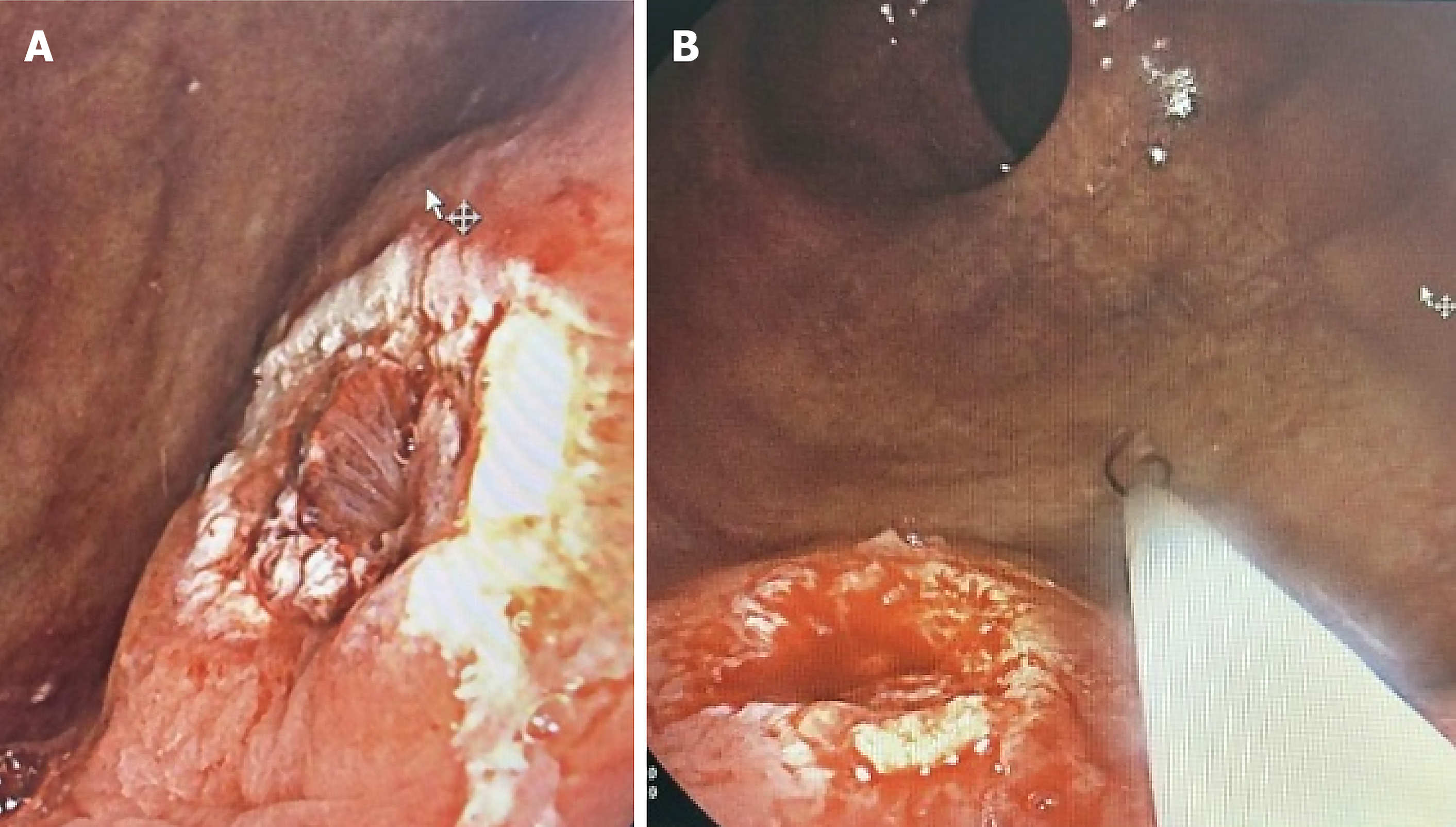
Figure 2 Successful endoscopic polypectomy using hot snare technique, with no bleeding observed at the resection site.
A: Lateral view of the polyp resection; B: Frontal view of the polyp resection.
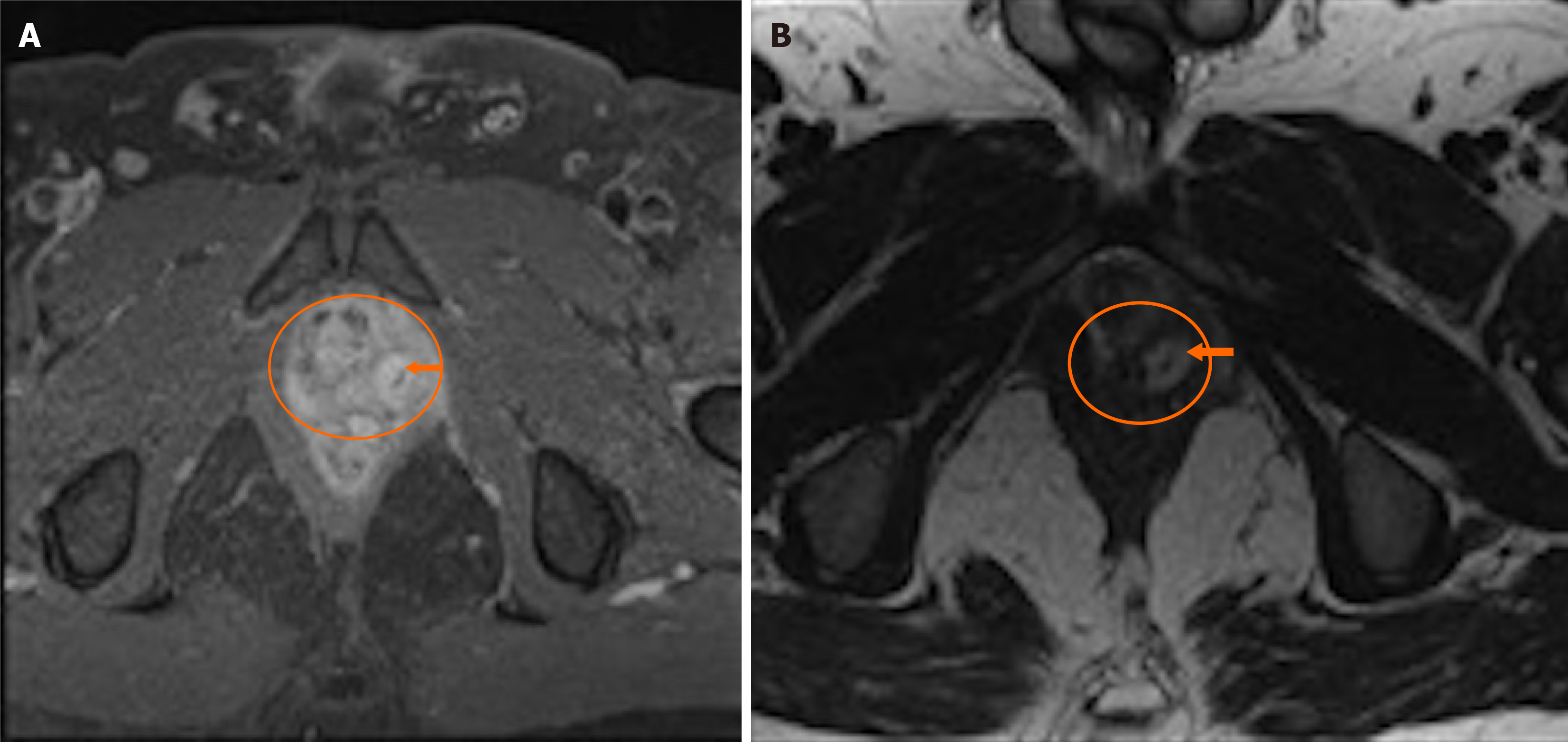
Figure 3 Intramural abscess and collection between the left iliococcygeal muscle and left prostatic lobe with thickened wall and peripheral enhancement post-contrast, along with a 34 mm linear hypodense lesion within the abscess (Foreign body).
A: T1 sequence of magnetic resonance imaging; B: T2 sequence of magnetic resonance imaging.
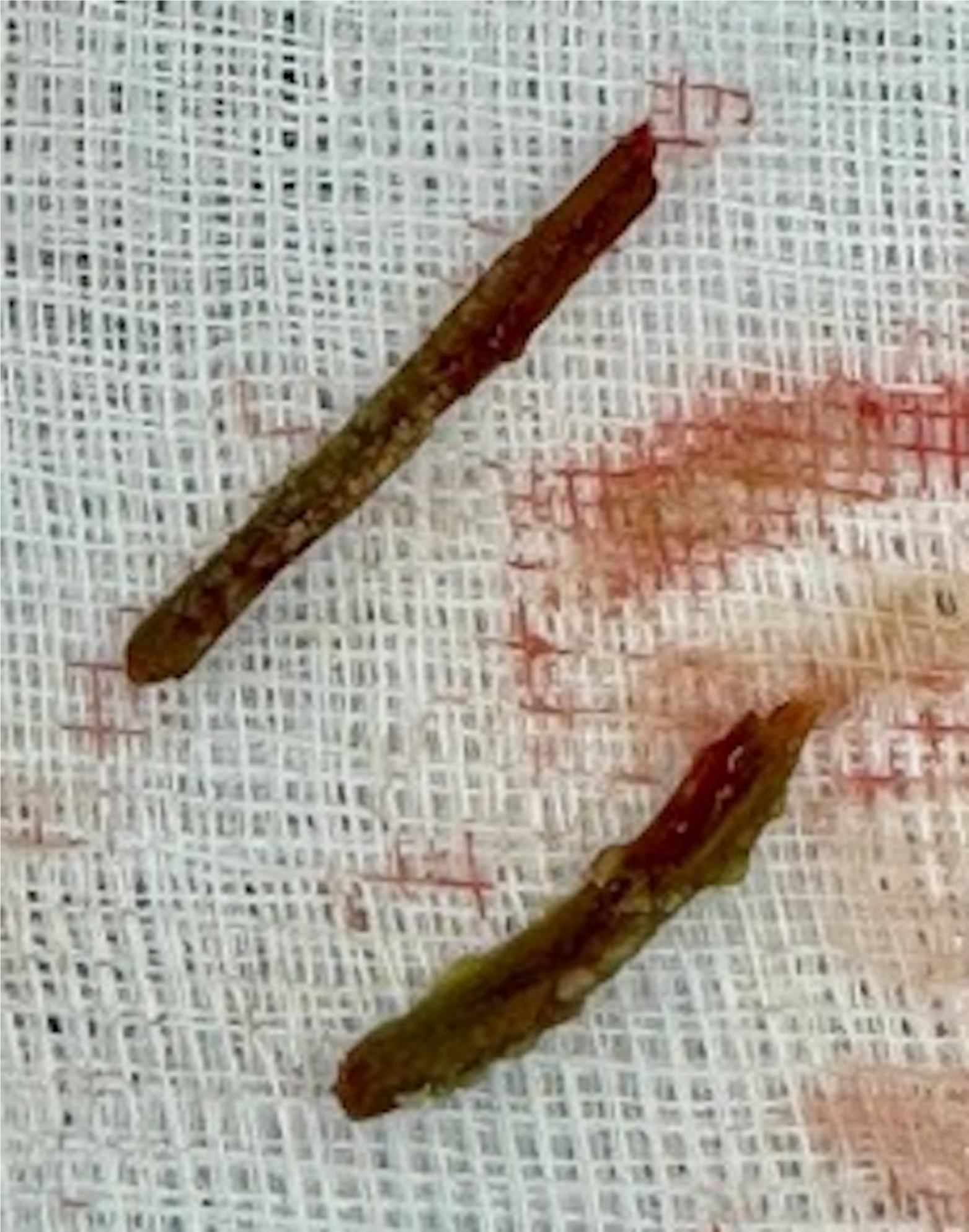
Figure 4
Bone fragments extracted during surgical resection.
- Citation: Martínez-Hincapie CI, González-Arroyave D, Ardila CM. Rectal abscess secondary to foreign body insertion: A case report. World J Clin Cases 2025; 13(18): 103438
- URL: https://www.wjgnet.com/2307-8960/full/v13/i18/103438.htm
- DOI: https://dx.doi.org/10.12998/wjcc.v13.i18.103438


