Copyright
©The Author(s) 2025.
World J Clin Cases. Jun 26, 2025; 13(18): 103055
Published online Jun 26, 2025. doi: 10.12998/wjcc.v13.i18.103055
Published online Jun 26, 2025. doi: 10.12998/wjcc.v13.i18.103055
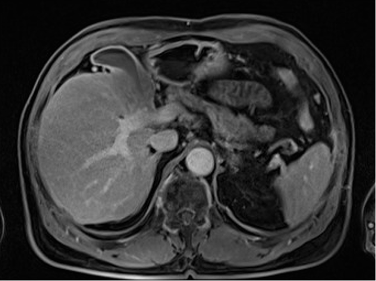
Figure 1 Magnetic resonance imaging of gallbladder.
Prostate magnetic resonance imaging depicted the gallbladder wall thickening (10 mm) with a low-intensity relative to the liver parenchyma on the T1-weighted.
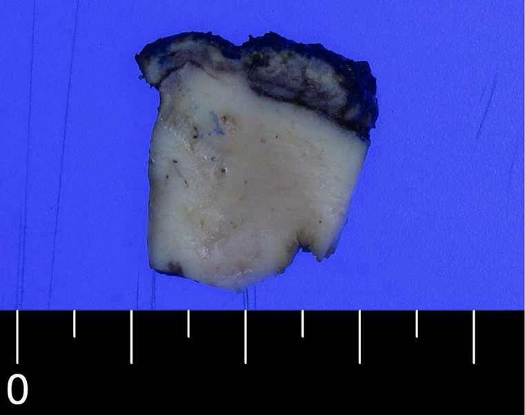
Figure 2 Macroscopic image of gallbladder.
The gallbladder wall appeared to be broadly thickened by approximately 1 cm.
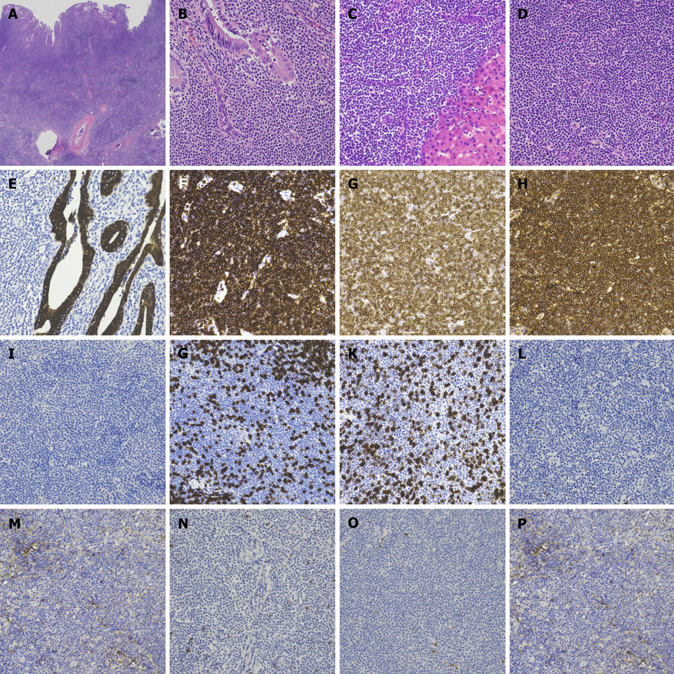
Figure 3 Histologic findings of mucosa associated lymphoid tissue lymphoma of the gallbladder.
A: Lymphoid tumor cells infiltrate whole layers of the gallbladder wall; B-C: Tumor cells infiltrate around the gallbladder mucosa (B) and liver parenchyma (C); D: Diffusely infiltrating tumor cells are small sized with inconspicuous nucleoli; E: Tumor cells infiltrate between cytokeratin-positive gallbladder mucosa, but do not form a lymphoepithelial lesion; F-H: Tumor cells are positive for CD20 (F), BCL2 (G), and Kappa light chain (H); I-P: Tumor cells are negative for TdT (I), CD3 (J), CD5 (K), CD10 (L), BCL6 (M), CD23 (N), cyclin D1 (O), and Lambda light chain (P). Original magnification: A: × 40; B-P: × 400.
Figure 4 Abdomen contrast-enhanced computed tomography imaging.
A: Thickened gallbladder wall (10 mm) was observed with homogeneous enhancement shown on the arterial; B: Thickened gallbladder wall (10 mm) was observed with homogeneous enhancement shown on the portal; C: Thickened gallbladder wall (10 mm) was observed with homogeneous enhancement shown on the delayed phases. Also, contrast-enhanced computed tomography showed laminar enhancement on the liver bed, which indicated invasion of the hepatic bed.
- Citation: Lee MR, Jang KY, Yang JD. Primary mucosa associated lymphoid tissue lymphoma of the gallbladder: A case report and review of literature. World J Clin Cases 2025; 13(18): 103055
- URL: https://www.wjgnet.com/2307-8960/full/v13/i18/103055.htm
- DOI: https://dx.doi.org/10.12998/wjcc.v13.i18.103055


