Copyright
©The Author(s) 2025.
World J Clin Cases. Jun 6, 2025; 13(16): 103373
Published online Jun 6, 2025. doi: 10.12998/wjcc.v13.i16.103373
Published online Jun 6, 2025. doi: 10.12998/wjcc.v13.i16.103373
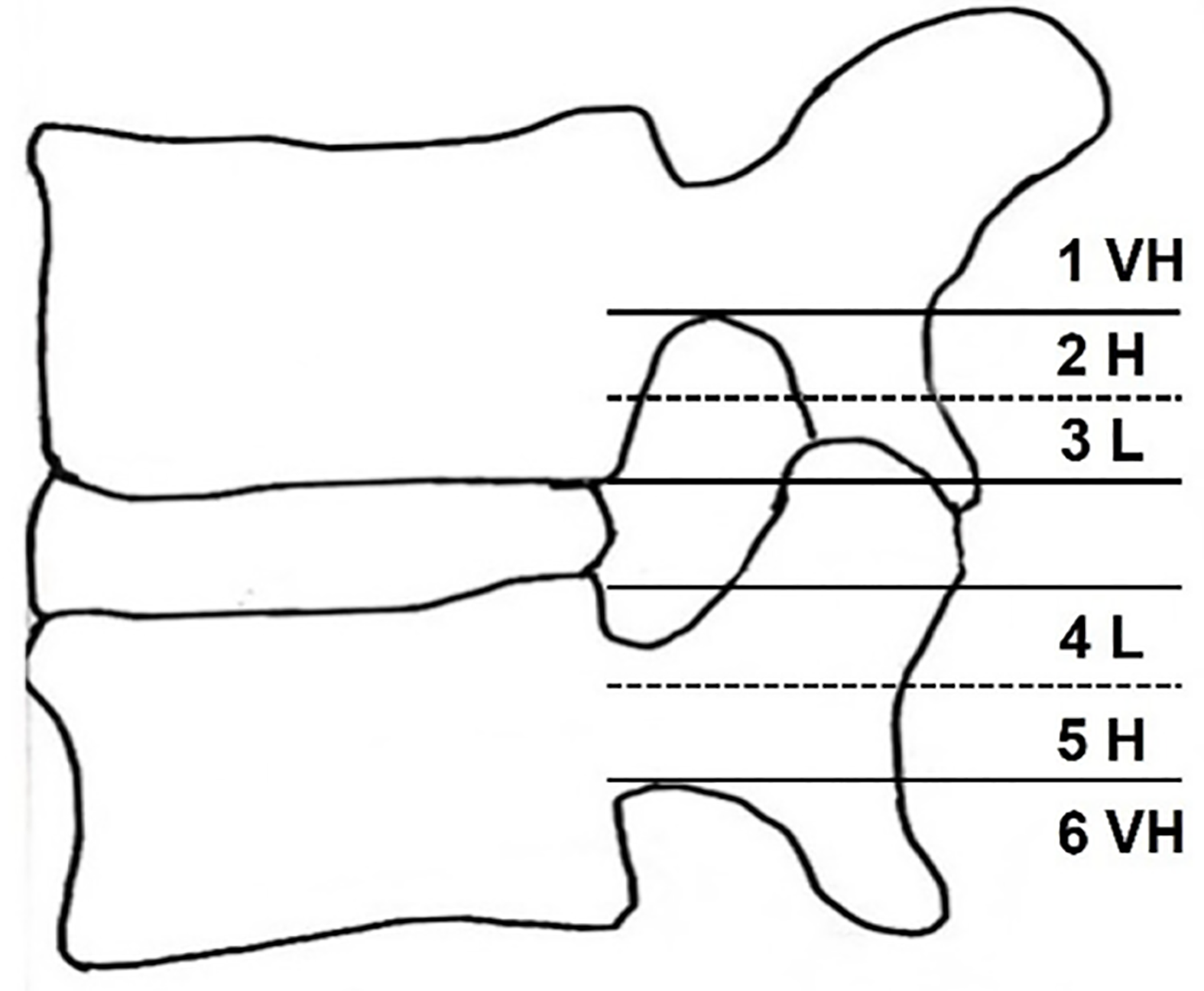
Figure 1 Schematic illustration of migrated lumbar disc grades.
A schematic illustration depicts the six grades of migrated lumbar discs in the sagittal plane, highlighting the varying degrees of migration: Very high (VH), high (H), and low (L).
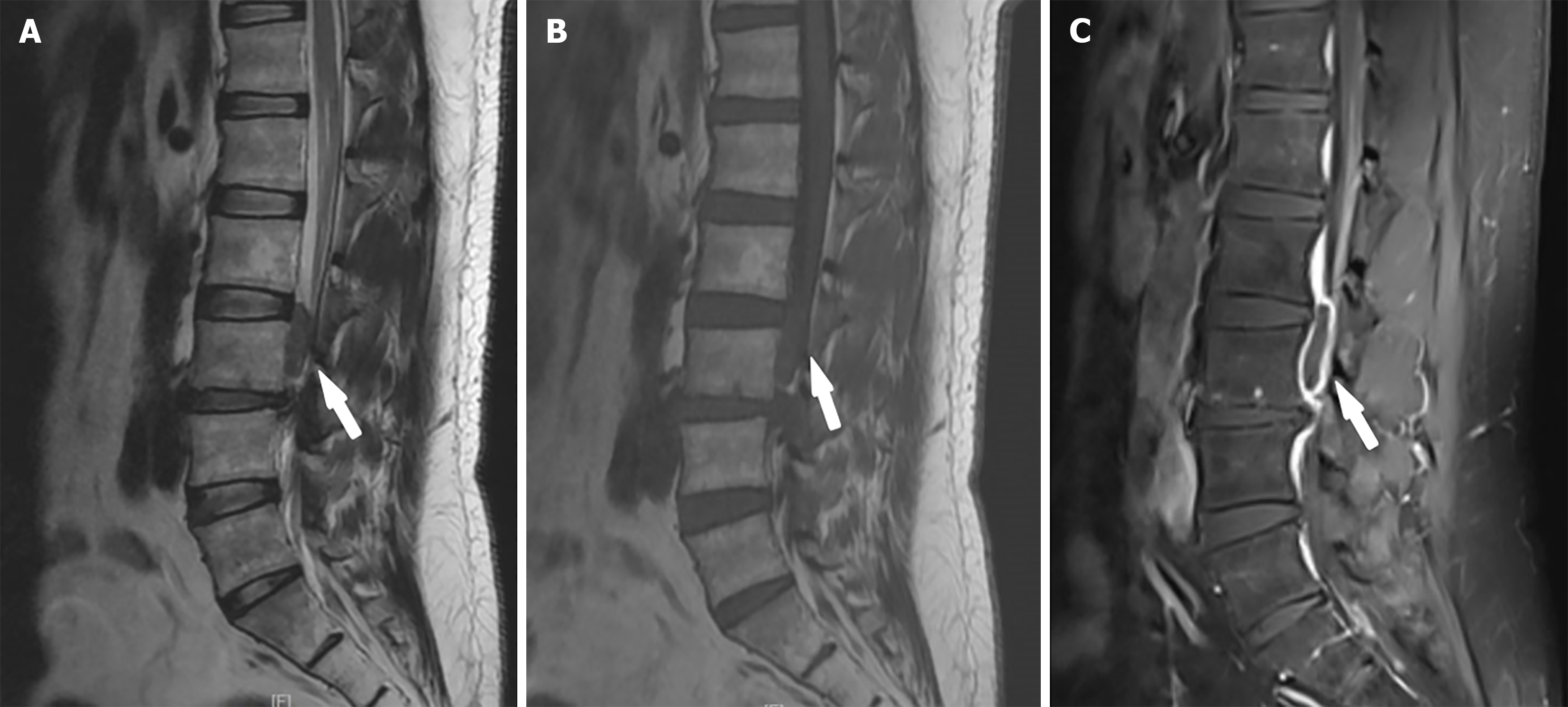
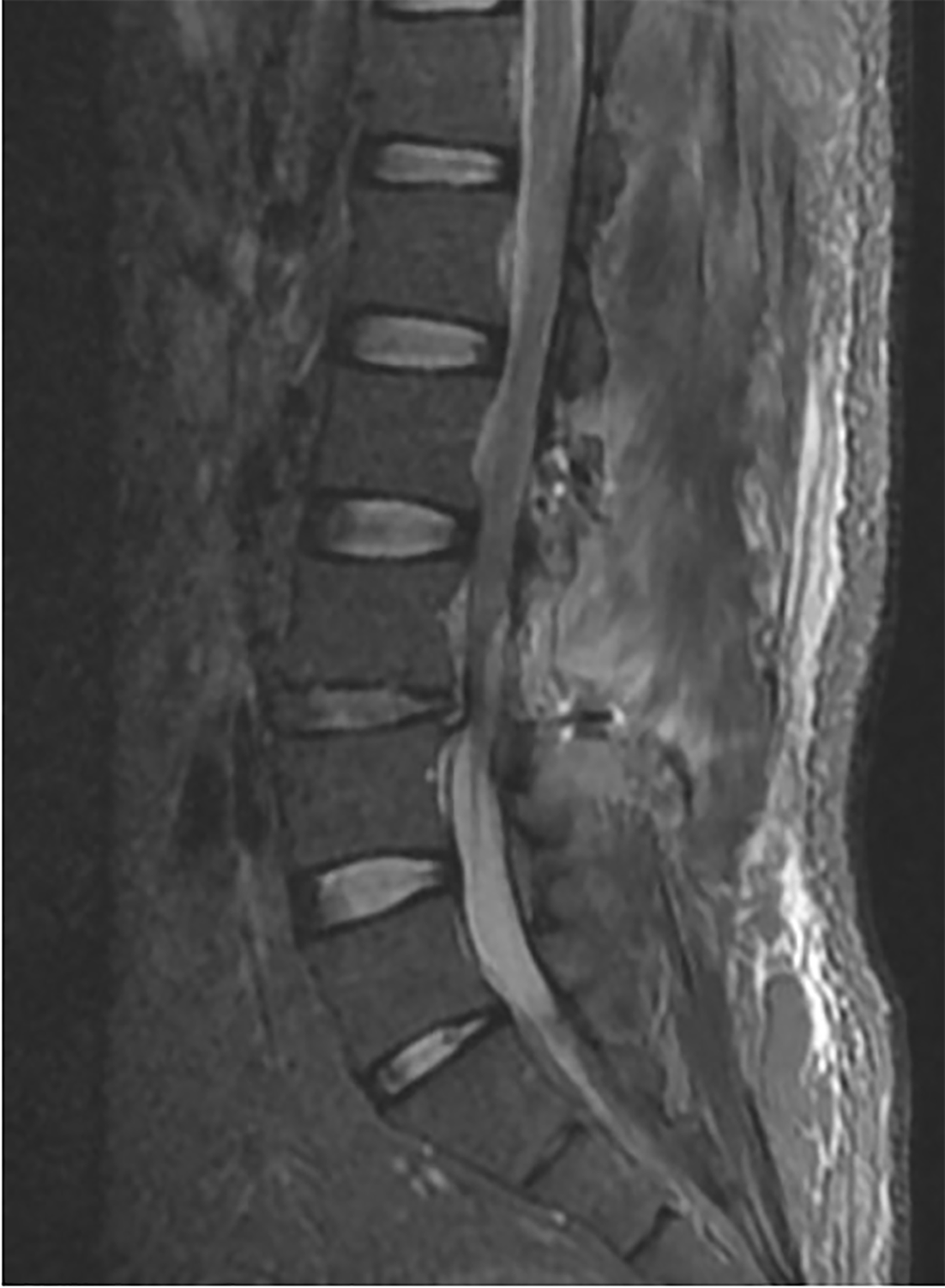
Figure 2 Lumbar spine sagittal weighted magnetic resonance imaging.
A: Sagittal T2-weighted magnetic resonance imaging (MRI) of the lumbar spine. The sagittal T2-weighted MRI scan revealed a significant reduction in the height of the L3-L4 intervertebral disc. A short T2 signal lesion is located posterior to the third lumbar vertebral body, as indicated by the arrow; B: Sagittal T1-weighted MRI of the lumbar spine. A long T1 signal lesion is located posterior to the third lumbar vertebral body, as indicated by the arrow; C: Sagittal T1-weighted enhanced MRI of the lumbar spine. In the sagittal MRI enhancement sequence, notable abnormalities were detected in the epidural elliptical signal located at the level of the third lumbar vertebra, as indicated by the arrow. These abnormalities were characterized by a prominent pattern of peripheral linear enhancement along the signal's edge, whereas the central region remained devoid of such enhancement.
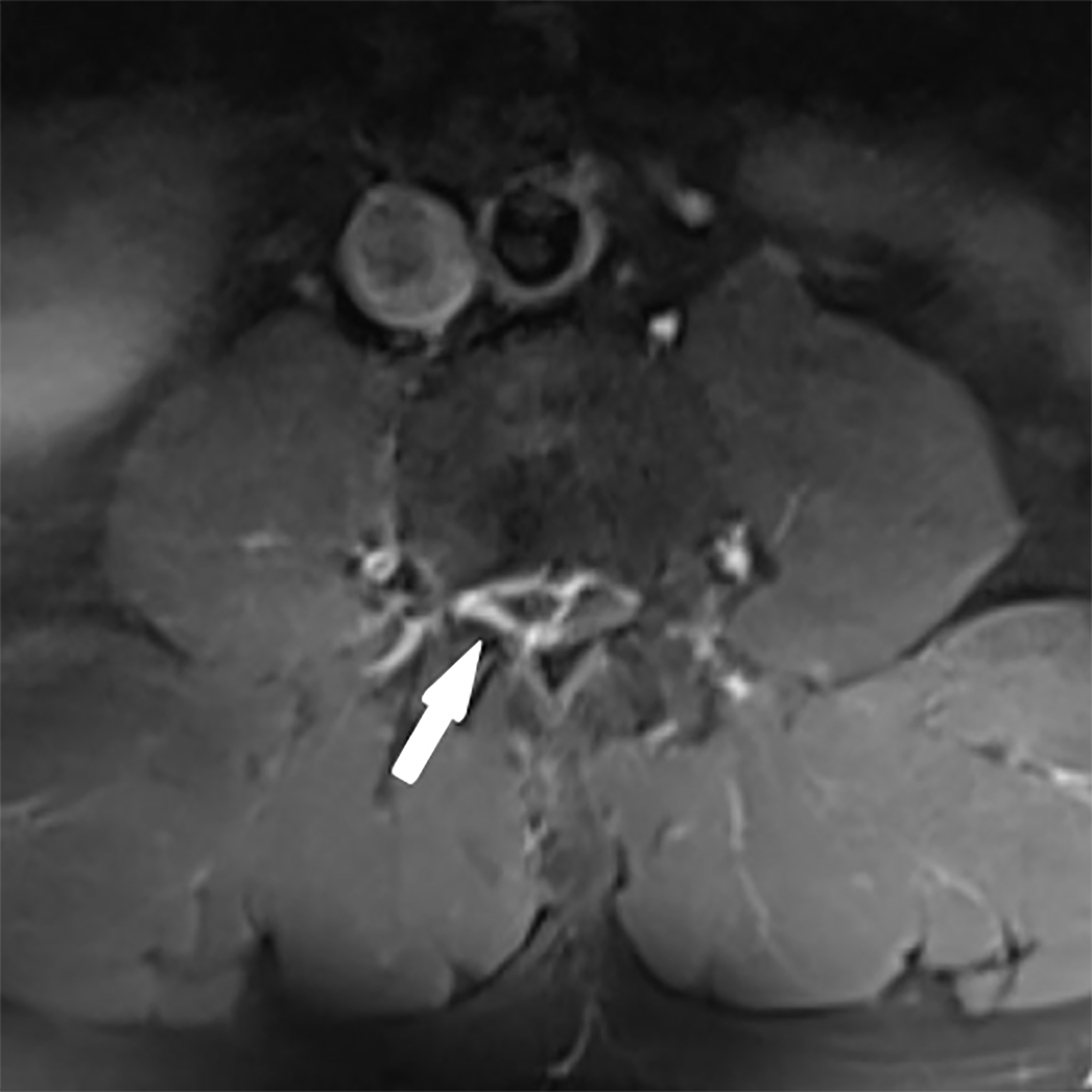
Figure 3 Axial T1-weighted enhanced magnetic resonance imaging of the lumbar spine.
Axial enhanced magnetic resonance imaging demonstrated a circumferentially enhanced lesion compressing the dural sac, resulting in its leftward deviation, as indicated by the arrow.
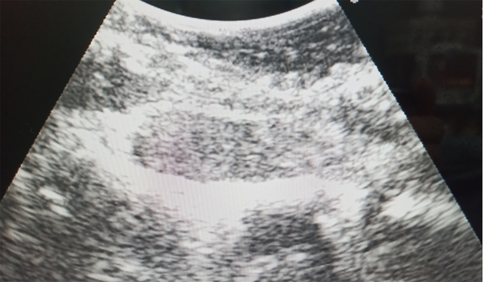
Figure 4 Intraoperative Ultrasonography for Localization of the Lesion Intraoperative ultrasonography precisely localized the lesion within the third vertebral body plane, situated along the ventral aspect of the spinal canal.
The lesion exhibited an elliptical shape and displayed a low signal echo, indicating its distinct characteristics.
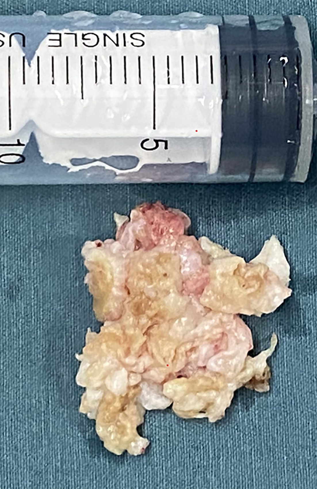
Figure 5 Pathological tissue analysis.
The observed pathological tissue exhibited a slightly yellowish color, with a brittle yet resilient texture, and lacked blood supply.
Figure 6 Postoperative sagittal T2-weighted magnetic resonance imaging of the lumbar spine.
The post-operative sagittal T2-weighted magnetic resonance imaging revealed repositioning of the dural sac at the level of the third lumbar vertebra.
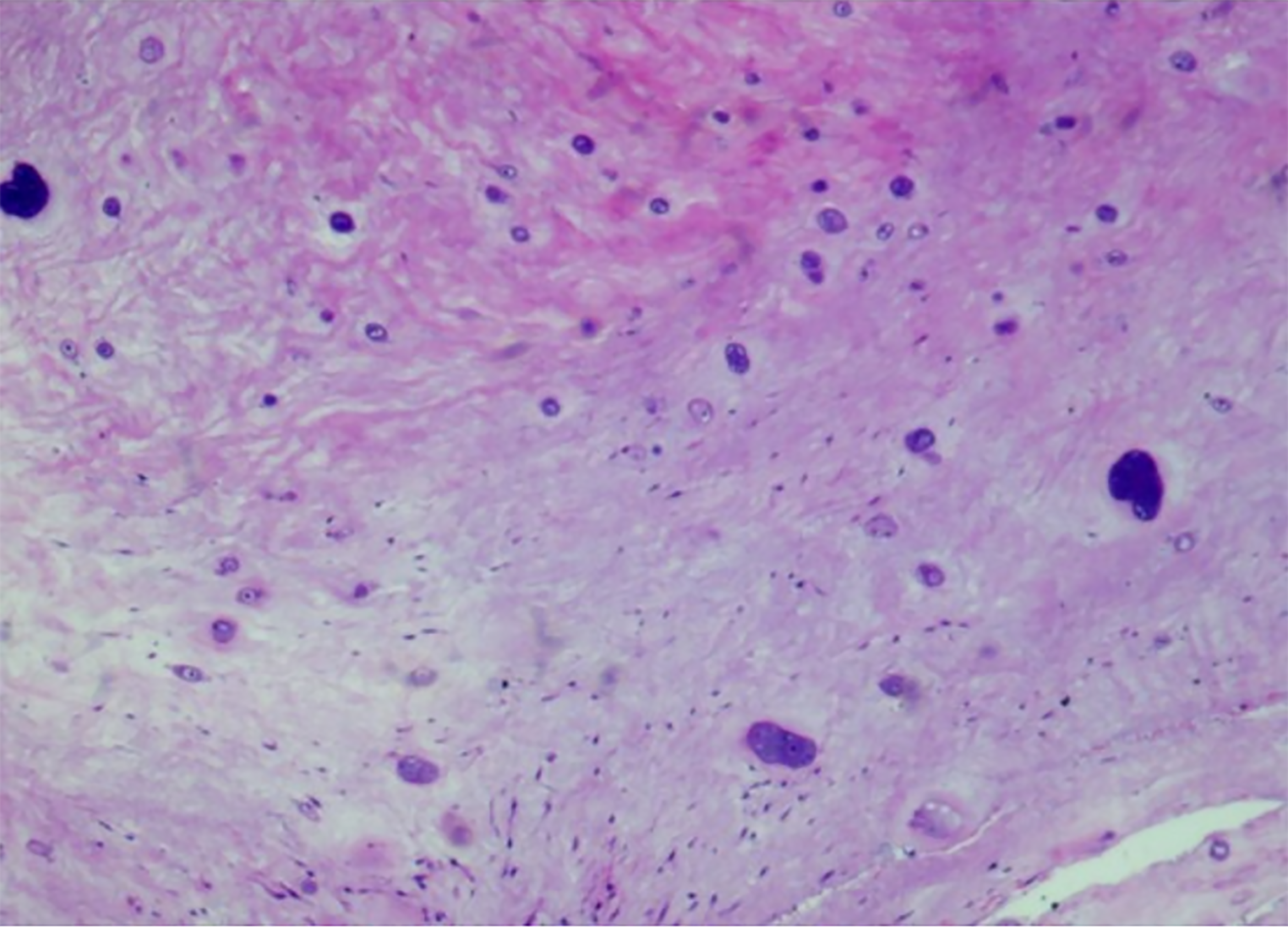
Figure 7 Postoperative pathological analysis of intervertebral disc tissue.
Postoperative pathological analysis revealed degenerated nucleus pulposus tissue originating from the intervertebral disc.
- Citation: Zhou JG. Diagnosis and surgical challenges of extremely severe head and lumbar disc herniation in young patients: A case report. World J Clin Cases 2025; 13(16): 103373
- URL: https://www.wjgnet.com/2307-8960/full/v13/i16/103373.htm
- DOI: https://dx.doi.org/10.12998/wjcc.v13.i16.103373


