Copyright
©The Author(s) 2025.
World J Clin Cases. May 26, 2025; 13(15): 99212
Published online May 26, 2025. doi: 10.12998/wjcc.v13.i15.99212
Published online May 26, 2025. doi: 10.12998/wjcc.v13.i15.99212
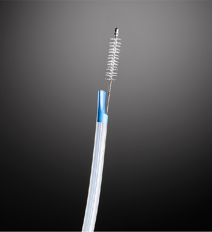
Figure 1
Close-up photo of the biliary cytology brush used in this study.
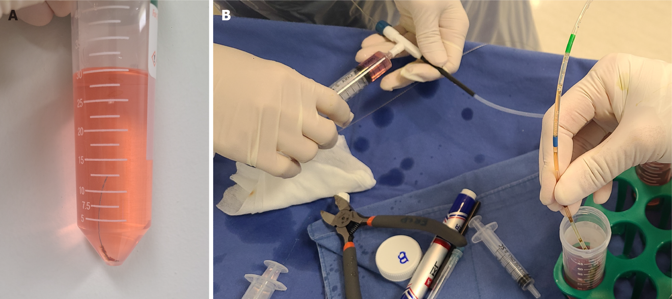
Figure 2 Preparation of the specimens.
A: Brush-wash specimens: The brush head was cut off the wire, placed in the medium, and centrifuged at 3000 r/m for 1 minute; B: Sheath-rinse specimens: The contents of the sheath were rinsed into the medium using a 10-cc syringe, and the specimens were centrifuged at 3000 r/m for 1 minute to isolate the cell pellet.
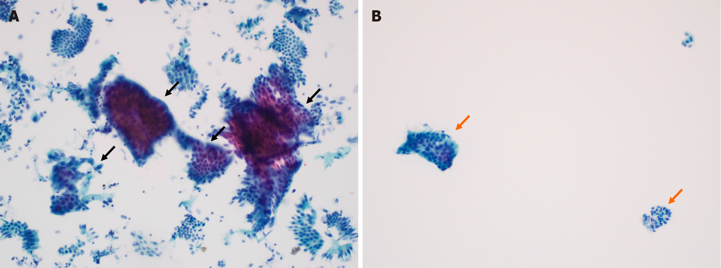
Figure 3 Representative examples of cytological specimens.
A: A brush specimen showing only a few medium and small clusters of biliary epithelial cells (black arrows) (papanicolaou stain, magnification 20 ×); B: A sheath-rinse specimen showing several large (> 50 cells) irregular clusters of malignant cells as well as medium (6-49 cells) and small (2-5 cells) clusters of biliary epithelial cells (orange arrows).
- Citation: So H, Jang SI, Ko SW, Yoon SB, Lee YS, Bang S, Kim M, Choi HJ. Effect of brush rinse on the diagnostic accuracy of biliary stricture evaluation: A multicenter trial. World J Clin Cases 2025; 13(15): 99212
- URL: https://www.wjgnet.com/2307-8960/full/v13/i15/99212.htm
- DOI: https://dx.doi.org/10.12998/wjcc.v13.i15.99212


