Copyright
©The Author(s) 2025.
World J Clin Cases. May 26, 2025; 13(15): 98608
Published online May 26, 2025. doi: 10.12998/wjcc.v13.i15.98608
Published online May 26, 2025. doi: 10.12998/wjcc.v13.i15.98608
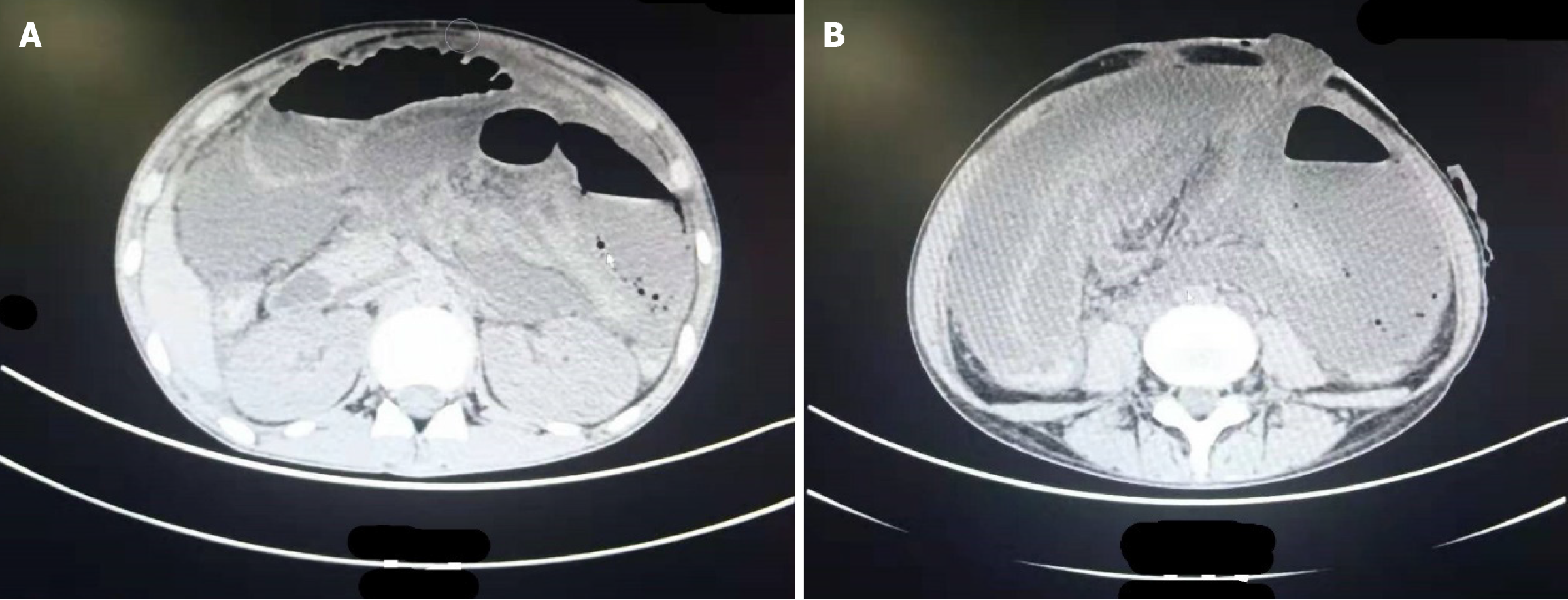
Figure 1 Computed tomography image of the abdominal cavity after an abdominal cocoon leading to stoma occlusion.
A and B: There was fluid accumulation and dilation caused by gas accumulation and obstruction in the intestinal lumen. There was multiple air-liquid levels in different planes.
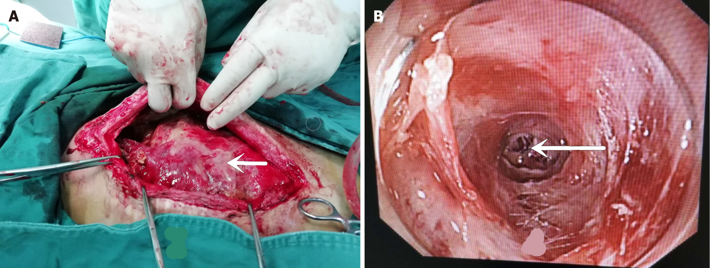
Figure 2 Stoma occlusion and adhesion of the intestine caused by abdominal cocoons.
A: Arrows indicate covered pus; B: The arrow indicates the blind end of the proximal small bowel occlusion.
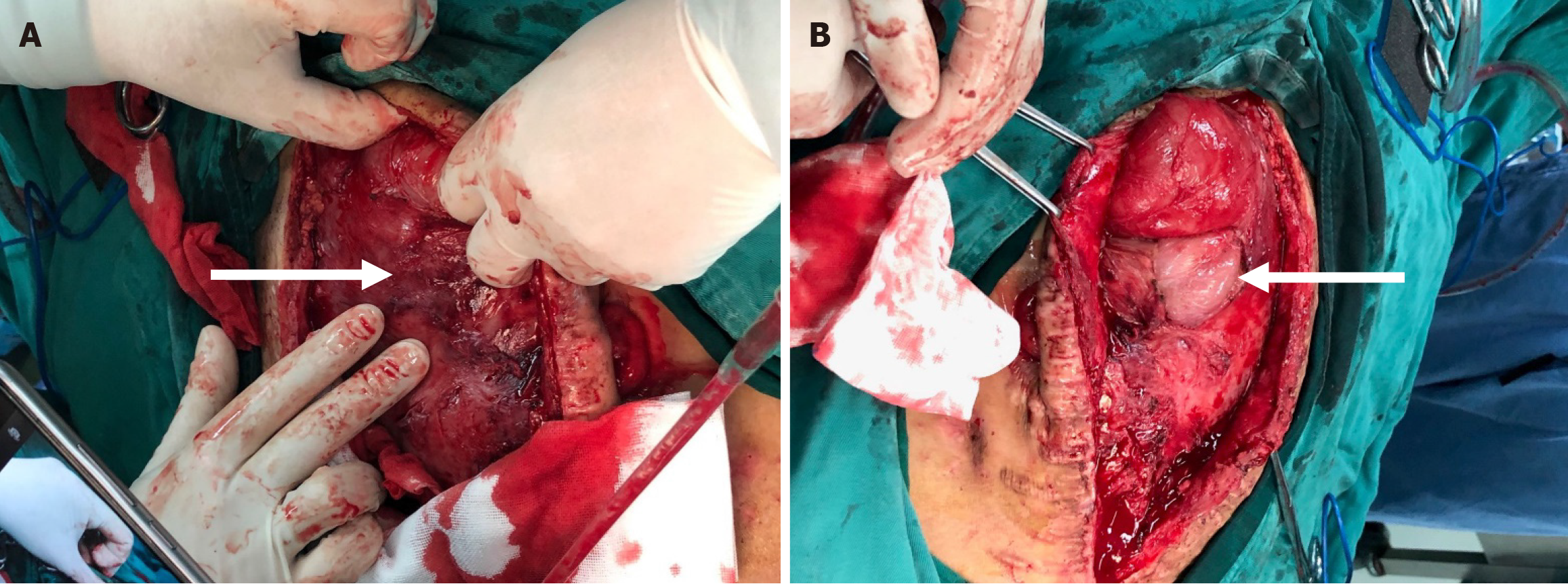
Figure 3 Fibrous membranes on the surface of the intestinal canal observed during abdominal cocoon surgery.
A: All organs in the abdominal cavity were covered with dense fibrous tissue of varying thickness (up to 2.0 cm), with thicker pelvic and lateral abdominal walls; B: After the cocoon-like membrane-like material was removed, the underlying intestinal tissue boundaries were clearer, and the adhesion between the intestinal tubes was not dense.
- Citation: Xu R, Sun LX, Chen Y, Ding C, Zhang M, Chen TF, Kong LY. Stoma occlusion caused by abdominal cocoon after abdominal abscess surgery: A case report. World J Clin Cases 2025; 13(15): 98608
- URL: https://www.wjgnet.com/2307-8960/full/v13/i15/98608.htm
- DOI: https://dx.doi.org/10.12998/wjcc.v13.i15.98608


